
Figures & data
Figure 1 The technique for intracanalicular implantation of the insert is shown. Following dilation of the lower canaliculus, the area is dried with a Weck-Cel® sponge. The insert is then carefully inserted into the lower canaliculus. Following insertion and contact with the tear film, the insert expands and conforms to the canalicular anatomy.
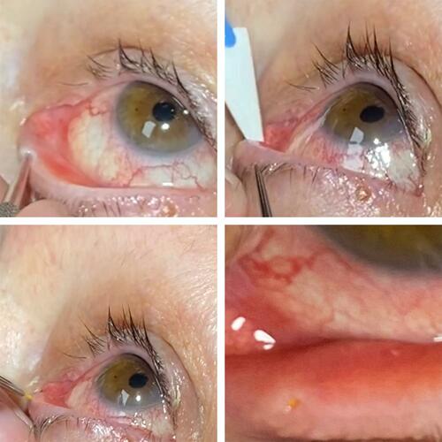
Table 1 Demographic and Clinical Characteristics
Table 2 Summary of the Presence of Anterior Chamber Inflammation at Each Time Point