
Figures & data
Table 1 Number and Percentage Distribution of Different Ophthalmological Diagnoses Among the Studied Patients
Table 2 Patients’ Characteristics According to Their Ophthalmological Diagnosis
Figure 1 Male patients 57 years old. He came to the ophthalmology clinic 1 months after recovery from COVID-19 infection, complaining of sudden diminution of vision. His optic disc was swollen and edematous with engorged vessels. His macular OCT scans show cystoid macular edema (CME) continuous with the disc edema with presence of neurosensory detachment (NSD).
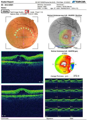
Figure 2 Male 52 year old diabetic patient just recovered from COVID presented with complete orbital apex syndrome (A and B). T2 FAT SAT MRI bilateral congested superior ophthalmic veins; red arrows (C). Coronal CT scan orbit and sinuses showing massive bone destruction red star (D).
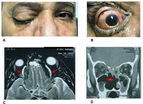
Table 3 COVID-19 Disease Manifestations Among Different Ophthalmological Implications