Figures & data
Table 1 Demographic Characteristics
Table 2 Clinical Characteristics, Treatment, and Follow-Up
Figure 1 (A and B) Slit lamp photography demonstrates thickening of lid margins, follicular reaction, and diffuse conjunctival injection. (C and D) With fluorescein instillation, negative staining reveals bulbar conjunctival follicular elevation.
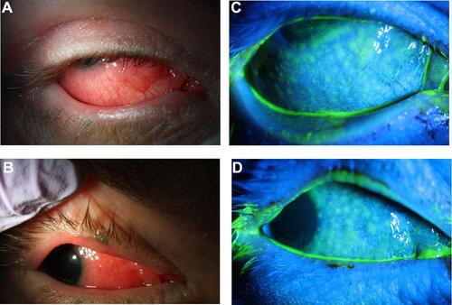
Figure 2 Right eye (left) and left eye (right) slit lamp photographs demonstrating superior conjunctival injection and limbal follicles.
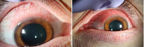
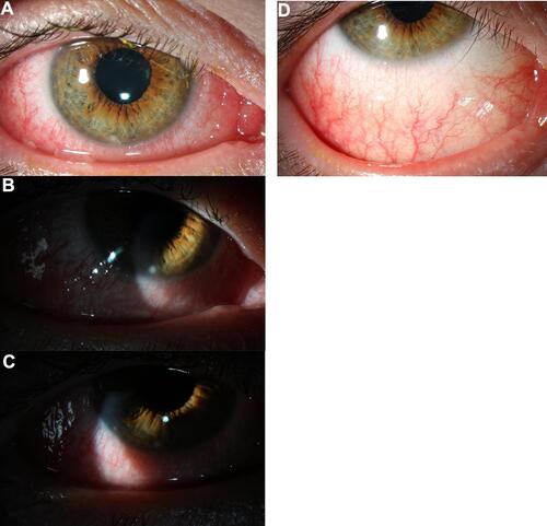
Figure 3 (A–C) Slit lamp photographs demonstrate diffuse conjunctival injection and 2 inferior marginal stromal infiltrates, at 6 o’clock and 8 o’clock. (D) After 1 week of treatment with topical fluorometholone 0.1% there is resolution of marginal stromal infiltrates.