Figures & data
Figure 1 Intrascleral knotless zigzag suture fixation of Akreos AO60 foldable intraocular lens (IOL) technique. (A) Conjunctival peritomy at 3 and 9 o’clock to expose the sclera. (B) Limbus corneal incision at 12 o’clock. (C) One haptic is externalized through the corneal incision. (D) A double thread 10-0 or 9-0 polypropylene suture is passed through the eyelet. (E) The needle is passed between the arms of the thread. (F and G) The needle is inserted through the corneal incision, passed behind the iris, and came out through the sclera at about 2 mm from the limbus. (H and J) The steps are repeated using the 180º away haptic and orienting the needle to the opposite side. (K) Centration of the IOL. (L–O) The suture is run through the partial thickness of the sclera in a zigzag pattern (4 intrascleral passages), and the thread is cut flush to the sclera without knotting (this procedure is performed in the nasal and temporal sides). (P) Conjunctival suture.
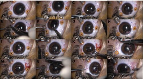
Figure 2 Postoperative slit-lamp photography of a patient that underwent intrascleral knotless zigzag suture fixation of Akreos AO60.

Table 1 Baseline Characteristics of Study Population
Table 2 Visual and Refractive Surgical Outcomes
Table 3 Perioperative Complications
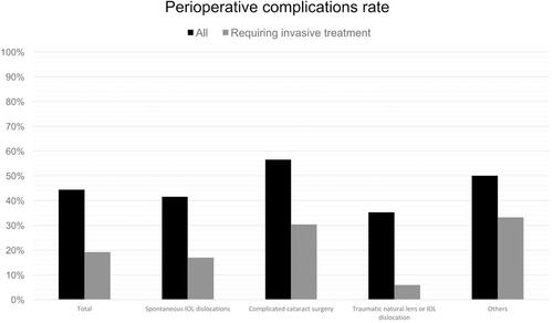
Figure 3 Perioperative complications rate.