Figures & data
Figure 1 Preoperative and postoperative photographs of patients with successful surgical outcomes. These patients demonstrated both excellent functional and symmetrical cosmetic outcomes with a favorable eyelid margin contour.
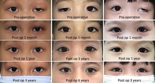
Figure 2 Surgical procedure; full-thickness maximal levator resection beyond Whitnall’s ligament, modified from Berke technique.Citation13 (A) eyelid crease skin incision. (B) orbital septum was opened to exposed preaponeurotic fat. (C) preaponeurotic fat was separated from the underlying levator aponeurosis. (D) superior 1/3 of tarsal plate was clearly exposed. (E) The Berke ptosis clamp was placed. (F) levator aponeurosis, Muller’s muscle, and conjunctiva were entirely detached from tarsus. (G) straight upward cuts to release the horns of the levator aponeurosis together with Whitnall’s ligament, and 1% lidocaine with 1:100,000 epinephrine was subconjunctivally injected. (H and I), palpebral conjunctiva was freed from the levator-Muller’s muscle flap, and reattached to superior boarder of tarsus. (J) the levator-Muller’s muscle flap was reinserted to the anterior surface (not below superior 1/3) of the tarsus (K) two additional fixation sutures were placed nasally and temporally for good eyelid contour. (L) the length of the redundant levator-Muller’s muscle flap was measured before resection. (M) the eyelid crease was created, and skin was closed.
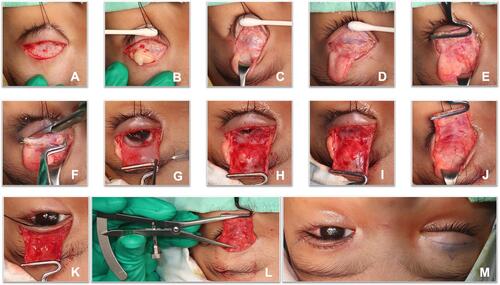
Table 1 Comparison of Demographic and Clinical Data Between Outcome Groups (31 Patients, 38 Eyelids)
Figure 3 Comparison of postoperative results (MRD1) between patients with successful and unsuccessful surgical outcomes, assessed over time.
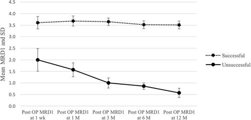
Table 2 Comparison of Postoperative Results (MRD1) Between Outcome Groups, Assessed Over Time
Table 3 Postoperative Functional Outcomes Assessed Over Time in 38 Eyelids
Table 4 Postoperative Symmetrical Cosmetic Outcomes in Patients with Unilateral Disease, Assessed Over Time in 24 Patients (MRD1 Compared Between Eyes)
Table 5 Complications of Maximal Levator Resection Beyond Whitnall’s Ligament in Patients Who Had Severe Simple Congenital Ptosis with Poor Levator Function
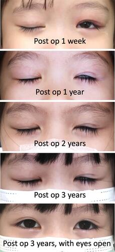
Figure 4 Photographs demonstrated improvement in postoperative lagophthalmos over time.