
Figures & data
Figure 1 Bicanalicular annular stent: (A) preparing the stent. (B) Pigtail probe was introduced into the lower punctum to get silicone tube from the wound. (C and D) Withdrawal of the pigtail probe from lower punctum. (E and F) This step repeated in the intact punctum. (G and H) Tying the proline 6\0 in 4 knots (I) Baring knots inside the stent then rotated to be inside the lacrimal sac. (J and K) Then canalicular edges were approximated using 6\0 vicryl sutures and repair lid laceration.
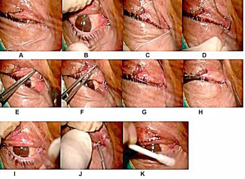
Figure 2 Bicanalicular nasal intubation: (A and B) A punctum dilator was used to enlarge the lacrimal puncti. (C) The proximal lacerated end was located. (D) One head was inserted into the lower canaliculus. (E) It was pulled from the inferior meatus of the nose. (F and G) The other end was placed into the upper canaliculus and pulled out from the nasal cavity by same technique. (H) Tie and trim ends of the tube in the nose. (I) The proximal and distal lacerated ends were subsequently anastomosed with 6\0 absorbable suture and Canalicular edges were approximated using 6\0 vicryl sutures.
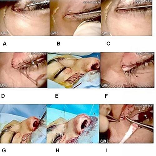
Table 1 Patients’ Characteristics
Figure 3 Bicanalicular annular stent; (A) preoperative. (B) postoperative.
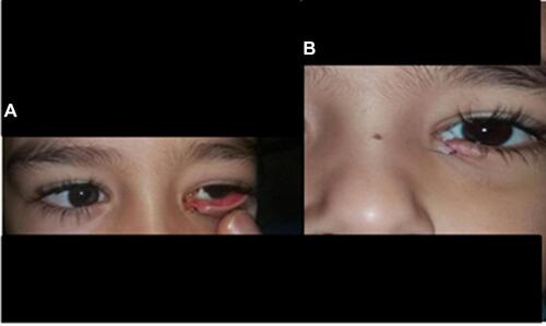
Figure 4 Bicanalicular nasal intubation; (A) preoperative. (B) postoperative.
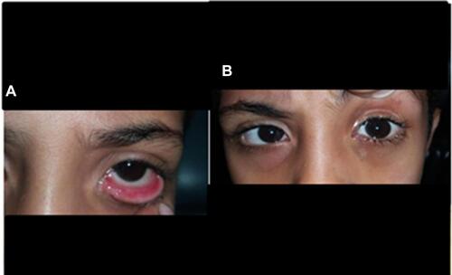
Figure 5 Premature extrusion of the tube after bicanalicular nasal intubation.
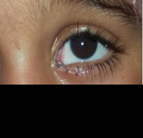
Figure 6 Punctal slit (cheese wiring) after bicanalicular nasal intubation.
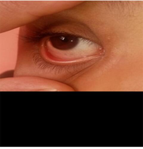
Table 2 Post-Operative Results