Figures & data
Figure 1 Workflow schema outlining the novel remote imaging approach used. (A) Both the on-site and off-site fellow communicate with each other as a part of the initial preparation stage. (B) The remote fellow then uses the institution approved remote computer to access an on-site workstation using the institutional user profile, (C) further connecting to and gaining full access to the advanced retinal imaging device. (D) Simultaneously, the on-site research fellow explains the study protocol and obtains a detailed written informed consent, (E) following which they accompany the patient to imaging room with the retinal imaging device and the on-site encrypted tablet computer device. (F) As the last step, the remote fellow uses the remote desktop to navigate the device for imaging and the remote encrypted tablet computer to communicate with the patient.
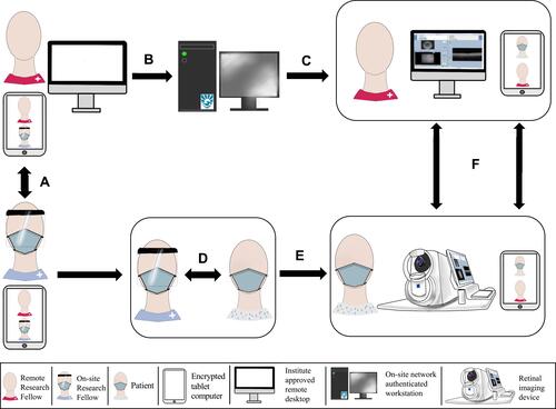
Figure 2 Representative images of patients being remotely imaged using widefield swept-source optical coherence tomography angiography device by an experienced research fellow via an institute approved remote desktop. The relevant communication was done via video conference call on an encrypted tablet device with hospital approved video call application. (Written patient consent obtained for being photographed here for the purpose of publication).
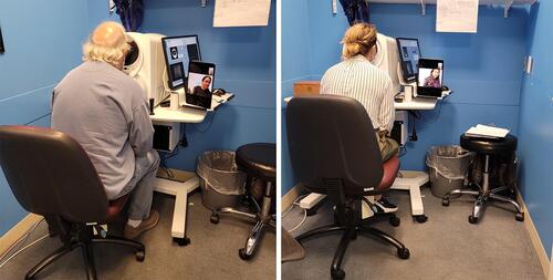
Table 1 Demographic Characteristics of Patients Imaged Remotely, Along with the Resultant Image Quality Metrics
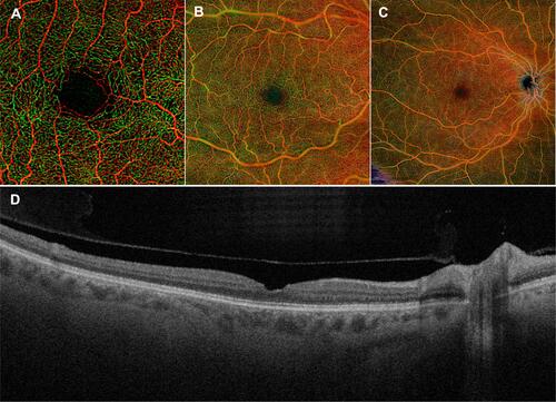
Figure 3 Representative Angio 3 mm x 3 mm (A), Angio 6 mm x 6 mm (B), 12 mm x 12 mm scans (C), and the Cube 800×800 B-scan (D) from the right eye of patient number 1 in , obtained via the remote imaging technique outlined.