
Figures & data
Table 1 The Preoperative Characteristics of the Three Studied Groups
Table 2 The Changes in MRD1 Measurements in the Three Studied Groups
Figure 1 Photos of a patient with severe left ptosis who was operated on using double sling technique. (A) Preoperative photo. (B) Photo taken in the early follow up period showing good initial postoperative outcome. (C) Photo taken 18 months after the surgery with good final postoperative outcome.
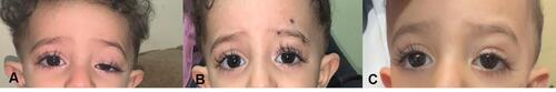
Table 3 Differences in the Surgical Outcomes Between the Studied Groups
Figure 2 Column chart showing the outcomes of surgery for the three studied groups assessed at the last follow up visit.
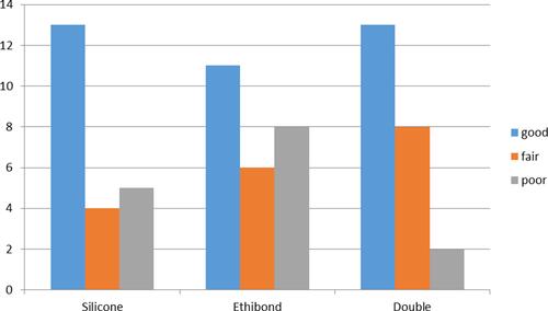
Table 4 Postoperative Symmetry in Bilateral Cases
Table 5 The Postoperative Main Complications in the Three Studied Groups
Table 6 The Results of Some Studies Conducted for the Use of Silicone Rod for Frontalis Suspension Surgery
Table 7 The Results of Some Studies Conducted for the Use of Ethibond for Frontalis Suspension Surgery