Figures & data
Figure 1 Anatomy, clinical picture, and histopathology of corneal epithelial basement membrane dystrophy (EBMD). EBMD occurs at the level of corneal epithelium and basement membrane (A). Clinically, it manifests as map-dot fingerprint opacities (B). Histology shows microcystic changes of the epithelial basement membrane (C).
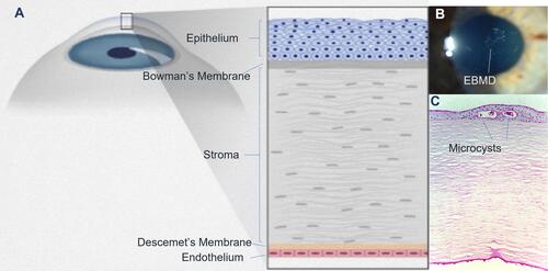
Figure 2 Images demonstrating clinically significant epithelial basement membrane dystrophy (EBMD). Slit lamp photography showing central focal anterior corneal opacities (A) Negative fluorescein staining can identify subtle EBMD and may extend beyond the visually involved area (B). Diagnosis can be confirmed with distorted Placido rings (C) and irregular mires on corneal topography (D).
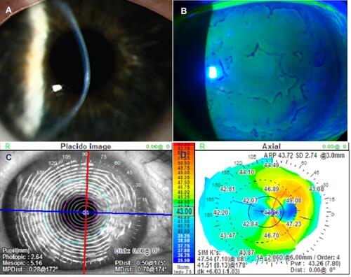
Figure 3 Slit lamp photograph at baseline, post-debridement, and post-cataract. Representative case in the study group showing clinically significant EBMD (A and B). One week after debridement and CAM placement, the ocular surface healed completely without positive or negative fluorescein staining (C and D), and remained stable after cataract surgery (E and F).
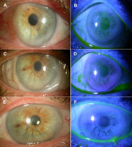
Table 1 Corneal Topograpy and Biometry Readings Before and 1 Month After Debridement in the Study and Control Groups
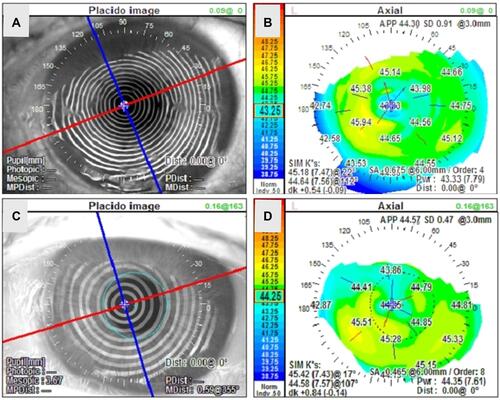
Figure 4 Representative topographical changes in the study group. At baseline (A and B) corneal topography showed marked irregular astigmatism and irregular Placido rings. One month after treatment (C and D), the astigmatism markedly reduced from 6D to 1D and the corneal curvature dramatically improved, while the Mean K changed from 43.3 D to 46.6 D along with concurrent ~ 30° axis deviation.
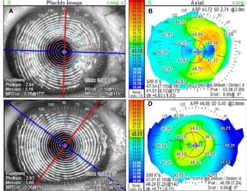
Figure 5 Representative topographical changes in the control group. similar changes were also observed in corneal topography and axis deviation at baseline (A and B) and 1 month after debridement (C and D).