Figures & data
Table 1 Clinical and Demographic Characteristics of Keratoconus Patients
Figure 1 Change in the rates of corneal topography and corneal tomography usage near the first visit over time. Corneal topography usage at the first visit stayed relatively constant from 2012 to 2019, whereas corneal tomography usage increased significantly since it was introduced in February 2015. Imaging near the first visit was defined as having either corneal topography or tomography done ±7 days from the first visit.
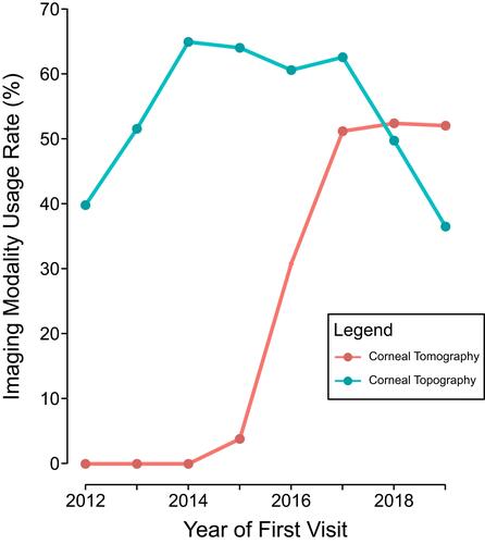
Table 2 Association of Imaging Modality and Intervention
Figure 2 Differences in interventions by the most recent visit for patients who did receive corneal tomography near the first visit compared to those who did not. (A) The percentage of patients with corneal collagen cross-linking (CXL) was significantly greater in those who did receive corneal tomography imaging near the first visit (P<0.001). (B) In contrast, the percentage of patients requiring another surgical intervention for keratoconus (defined as having ICRS, lamellar keratoplasty, or penetrating keratoplasty) was significantly lower in patients who did have corneal tomography near the first visit compared to patients without tomography (P=0.048). Statistics: Multivariate linear regression controlling for year of the first visit. *P<0.05, **P<0.001.
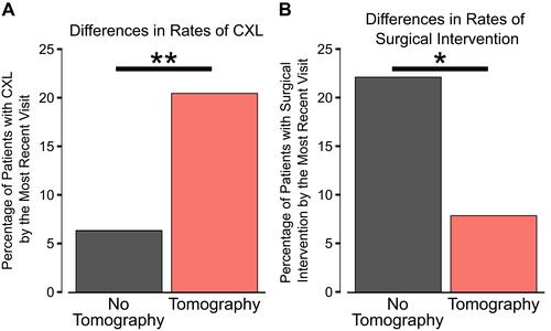
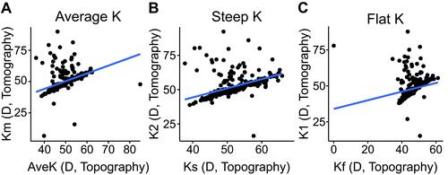
Figure 3 Correlation between keratometry measurements from corneal topography and corneal tomography for patients at their first visit. Patients with corneal topography and tomography within 30 days from each other were included (N=690 eyes). (A) The correlation between the average keratometry from corneal topography (AveK) and tomography (Km) had an adjusted R2 of 0.16 (P<0.001). (B) The adjusted R2 value for the correlation between the steep K from corneal topography (Ks) and tomography (K2) was 0.21 (P<0.001). (C) The adjusted R2 value for correlation between the flat K from corneal topography (Kf) and tomography (K1) was 0.04 (P<0.001). All K values are in diopters (D).