Figures & data

Figure 1 Diffuse vitreous opacification illuminated intraoperatively. Image courtesy of Retina Specialists of Alabama LLC (RSA).
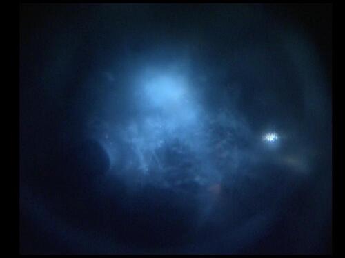
Figure 2 Fundus photograph showing a single, focal SVO in the central, posterior vitreous that was highly symptomatic and non-resolving. Image courtesy of RSA.
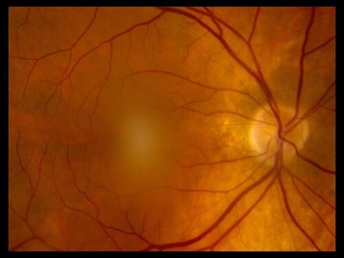
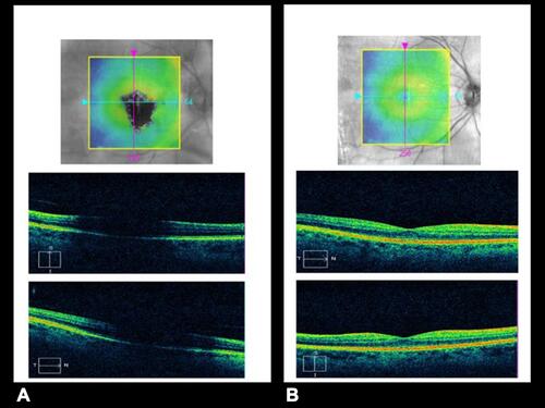
Figure 3 (A) Preoperative OCT image showing shadowing of the macula by the same SVO seen in , mimicking the recurrent SVO scotoma seen by the DVS patient. (B) Postoperative OCT image after VOV showing absent shadowing and a high-quality image through a clear visual axis. Images courtesy of RSA.