Figures & data
Figure 1 Fluorescein angiography. Case 1: macular edema without macular ischemia. Case 2: macular edema associated with ischemia of the macular region.
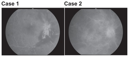
Figure 2 Changes in mean macular thickness (A–C) after intravitreal injection of triamcinolone acetonide in two patients with branch retinal vein occlusion and macular edema. (A) Case 1: mean macular thickness within the central 4 degree field is reduced after 3 and 6 months. Case 2: mean macular thickness within the central 4 degree field is reduced after 3 months, but increased at 6 months compared with at 3 months. (B) Case 1: mean macular thickness within the 10 degree field is reduced after 3 and 6 months. Case 2: mean macular thickness within the 10 degree field is reduced after 3 months, but increased at 6 months compared with at 3 months. (C) Case 1: mean macular thickness within the 20 degree field is reduced after 3 and 6 months. Case 2: mean macular thickness within the 20 degree field is reduced after 3 months, but increased at 6 months compared with at 3 months.
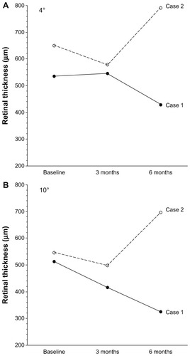
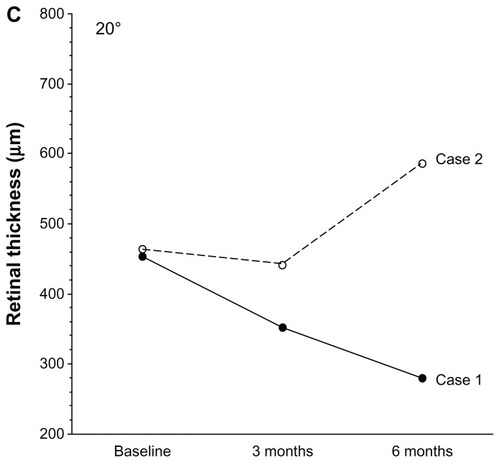
Figure 3 Changes in best-corrected visual acuity after intravitreal injection of triamcinolone acetonide in two patients with branch retinal vein occlusion and macular edema. Case 1: best-corrected visual acuity is improved after 3 and 6 months. Case 2: best-corrected visual acuity is not improved after 3 or 6 months.
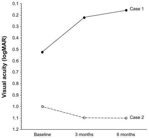
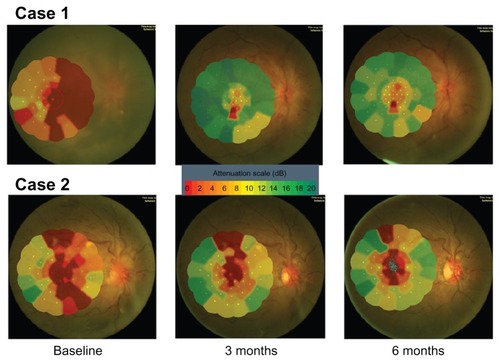
Figure 4 Changes in macular sensitivity (A–C) after intravitreal injection of triamcinolone acetonide in two patients with branch retinal vein occlusion and macular edema. (A) Case 1: mean macular sensitivity within the central 4 degree field is increased after 3 months, but decreased at 6 months compared with at 3 months. Case 2: mean macular sensitivity within the central 4 degree field is not improved after 3 or 6 months. (B) Case 1: mean macular sensitivity within the 10 degree field is increased after 3 months, but decreased at 6 months compared with at 3 months. Case 2: mean macular sensitivity within the 10 degree field is almost unchanged after 3 and 6 months. (C) Case 1: mean macular sensitivity within the 20 degree field is increased after 3 months, but decreased at 6 months compared with at 3 months. Case 2: mean macular sensitivity within the 20 degree field is almost unchanged after 3 and 6 months.
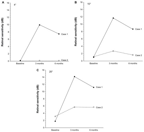
Figure 5 Macular sensitivity map obtained by microperimetry.