Figures & data
Figure 1 External photograph and axial CT scan of the same patient. External photograph of the patient showing right eye proptosis with periocular swelling and mild conjunctival congestion (A). CT scan, axial cut, of the same patient showing a large mass occupying the lateral quadrant of the orbit with erosion of the lateral wall and extension into the temporal fossa (B).
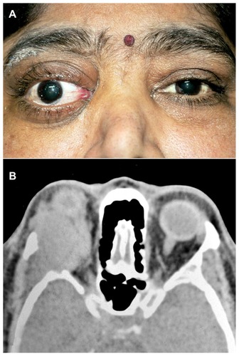
Figure 2 Microphotograph and immunohistochemical staining. Microphotograph showing atypical plasmacytoid tumors cells with abundant amphophilic cytoplasm and a large eccentric vesicular nucleus having a prominent nucleolus (HE × 400) (A). Immunohistochemical staining shows lack of immunoreactivity for CD20 (×400) (B). Strong membranous CD138 immunoreactivity (×400) (C). Strong Ki-67 immunoreactivity in almost all tumors cells (×400) (D).
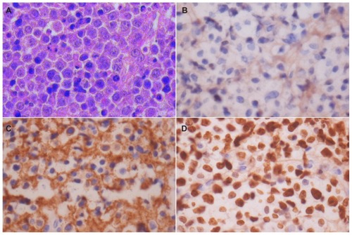
Figure 3 External photograph and CT scan of the patient showing gross proptosis of the left eye with severe conjunctival chemosis, and a large mass lesion in the superior orbit. External photograph of the patient showing gross proptosis of the left eye with severe conjunctival chemosis (A). CT scan with sagittal reconstruction showing a large mass lesion in the superior orbit with extension into the frontal sinus and intracranial space (B).
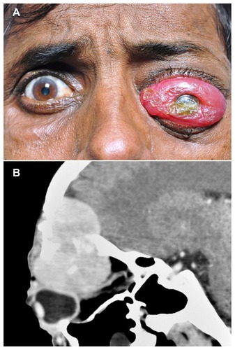
Figure 4 External photograph of the patient showing periocular edema with total ptosis, skin induration, and blisters, as well as lip edema and blisters. External photograph of the patient showing left periocular edema with total ptosis, skin induration, and blisters (A). External photograph showing upper lip edema with blisters (B). CT scan coronal cut of the same patient shows a diffuse ill-defined mass involving the entire superior orbit with superolateral bony erosion and extension into the temporal fossa (C). CT scan with sagittal reconstruction showing diffuse mass involving the entire superior quadrant up to the apex with indentation of the globe and optic nerve stretch (D).
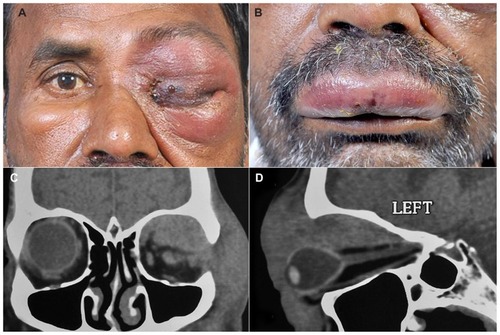
Table 1 Summary of clinical, radiological, and histopathologic features of ocular plasmablastic lymphomas reported in the literature
Table 2 Clinicopathological features of cases of plasmablastic lymphoma included in the present study