Figures & data
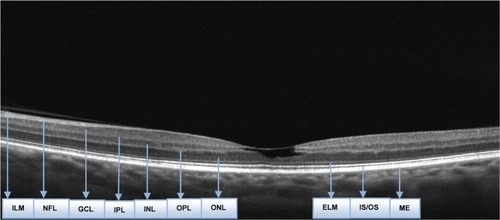
Figure 1 Optical Coherence Tomography of the right eye.
Abbreviations: ELM, External Limiting Membrane; GCL, Ganglion Cell Membrane; ILM, Internal Limiting Membrane; INL, Inner Nuclear Layer; IPL, Inner Plexiform Layer; IS/OS, Inner-Outer Segment Photoreceptors; NFL, Nerve Fiber Layer; OPL, Outer Plexiform Layer; ONL, Outer Nuclear Layer; PE, Retinal Pigment Epithelium.
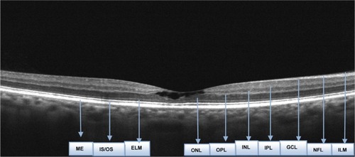
Figure 2 Optical Coherence Tomography of the left eye.
Abbreviations: ELM, External Limiting Membrane; GCL, Ganglion Cell Membrane; ILM, Internal Limiting Membrane; INL, Inner Nuclear Layer; IPL, Inner Plexiform Layer; IS/OS, Inner-Outer Segment Photoreceptors; NFL, Nerve Fiber Layer; OPL, Outer Plexiform Layer; ONL, Outer Nuclear Layer; PE, Retinal Pigment Epithelium.