Figures & data
Figure 1 (1a and b) Performing supraorbital and infraorbital nerve block using JBP 33 gauge 4 mm long nanoneedles. (2a and b) Jojoba anesthetic ointment consisting of 8% lidocaine and 25% jojoba is taken from a refrigerated jar and applied to the lid margin for 10 minutes. This may be repeated. (3) The probing set-up at the slit lamp with an assistant to steady the patient for good visualization. (4) View through the slit lamp microscope of probing upper (a) and lower (b) lid Meibomian glands. (5) Meibography guided probing using the Mediworks S390L WDR FireFly Digital Slit Lamp from Eyefficient (Aurora, Ohio), demonstrating a 4 mm probe within the central duct (a), a sterile MicroTube Stent within the central duct for retrieval of meibum (b) and the retrieved meibum inside the MicroTube removed from within the gland (c). Reproduced from Maskin SL, Alluri S. Meibography guided intraductal meibomian gland probing using real-time infrared video feed. Br J Ophthalmol. 2020;104(12):1676; with permission from BMJ Publishing Group Ltd.Citation21 (6) An alternative approach to obtaining a virgin sample of meibum by allowing the meibum to travel through the MicroTube Stent for collection and analysis. Arrow shows a drop of meibum at the distal end of the MicroTube Stent.Citation22
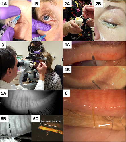
Table 1 Summary of Independent MGP Studies and Their Result
Figure 2 Probe findings form of a patient.
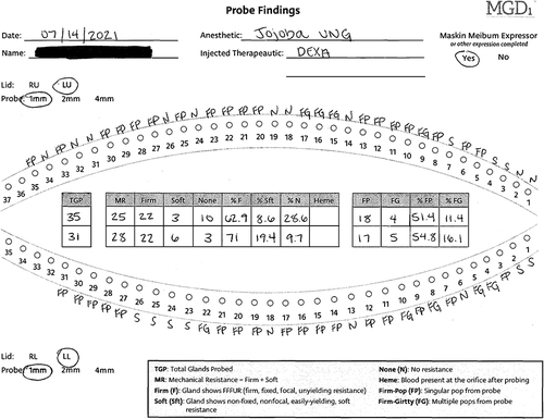
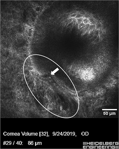
Figure 3 Confocal microscopy image of Meibomian gland distal duct showing disruption of the normally well demarcated external duct wall by fibrovascular tissue invasion. A prominent blood vessel is seen inside the oval. The disruption of the duct wall is indicated by the solid arrow showing a “step off”. This gland had not been probed. (Courtesy of SLM.).