Figures & data
Table 1 Patients’ characteristics
Figure 1 Types of hemangioma according to depth of involvement.
Notes: (A) Superficial left upper-eyelid hemangioma. (B) Subcutaneous left lower-eyelid hemangioma. (C) Combined superficial and subcutaneous hemangioma in the glabella.
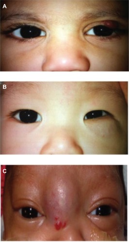
Table 2 Mean visual acuity, and sphere and cylinder refraction at first and last visits
Table 3 Modes of treatment used for periocular capillary hemangiomas in this series
Figure 2 Photograph of one case of capillary hemangioma that resolved spontaneously without treatment at (A) 4 months old upon first visit and at (B) 5 years old upon latest visit.
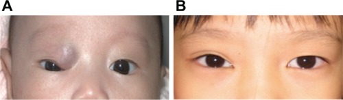
Figure 3 A 6-month-old female with combined superficial and deep hemangioma over the right superior orbit with inferior globe displacement.
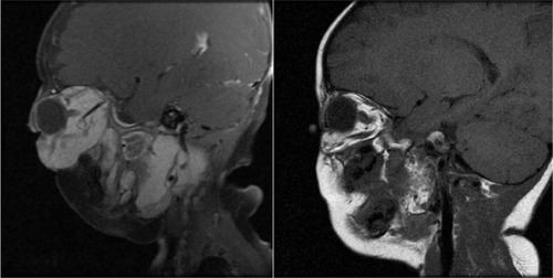
Abbreviation: MRI, magnetic resonance imaging.
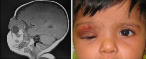
Figure 4 Sagittal MRI of a hemangioma involving the left orbit and pterygopalatine fossa before (left) and 1 year after treatment (right) with oral prednisolone.