Figures & data
Figure 1 (A) Scheimpflug image in a keratoconus patient. (B) Axial (power) map, anterior and posterior elevation maps, and global pachymetry map in a keratoconus patient.
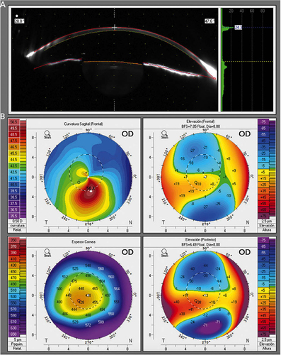
Figure 2 Schematic depiction of the Dresden protocol: removal of corneal epithelium, instillation of riboflavin solution, and application of a UV-A beam of 370 nm wavelength.
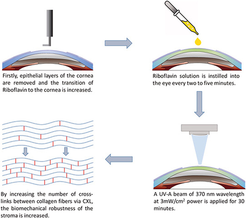
Figure 3 Intracorneal ring segments implanted within the cornea.
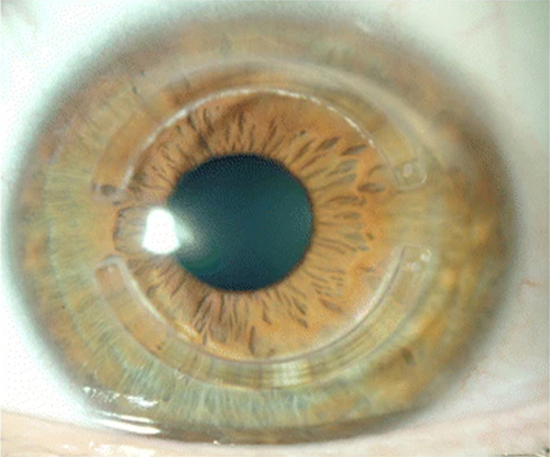
Figure 4 (A and B) Introduction of a CAIRS into an intrastromal channel. The arrows highlight the CAIRS it is being inserted (C and D) Two CAIRS within the intrastromal channel.
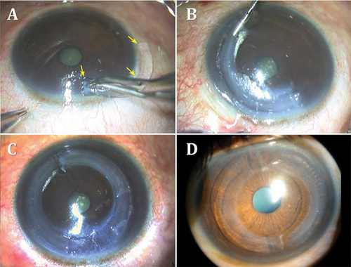
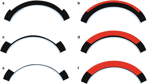
Figure 5 Schematic representation of the anterior lamellar keratoplasty technique with varying depths of recipient cornea removal and donor corneal transplantation. (a and b) One-third of the anterior cornea is removed and replaced with a similarly sized donor cornea. (c and d) Larger amount of anterior cornea is removed and replaced. (e and f) Deep anterior lamellar keratoplasty where corneal tissue is removed up to the bare Descemet membrane and donor cornea without Descemet membrane is transplanted.