Figures & data
Table 1 Review of Literature of DMEK Graft Rejection (Larger Case Series) of the Past 5 Years
Table 2 Review of Literature of DMEK Graft Rejection (Case Reports) of the Past 5 Years
Figure 1 Depicting the CONSORT flowchart of the systematic review.

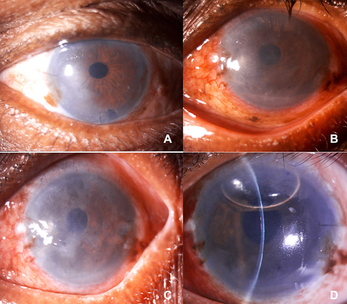
Figure 2 (A) Digital slit lamp image depicting a patient with pseudophakic bullous keratopathy planned for DMEK (B) Digital slit lamp image of a patient with Fuch’ Endothelial Corneal Dystrophy planned for DMEK (C) and (D) Digital slit lamp images of patients post-DMEK depicting a well opposed lenticule with relatively clear cornea.
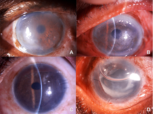
Figure 3 (A) Digital slit lamp image of a patient post DMEK, depicting stromal edema in the inferior one-third of cornea suggestive of early graft rejection.(B) Digital slit lamp image of a patient on a postoperative day 1, post-DMEK, depicting corneal haze with stromal edema suggestive of primary graft failure. (C) Digital slit lamp image of a patient on postoperative one month, post DMEK, depicting conjunctival congestion, corneal haze with stromal edema suggestive of primary graft failure. (D) Digital slit lamp image of a patient post DMEK following rebubbling, depicting clear cornea, well opposed lenticule with an air bubble occupying 1/3rd anterior chamber.