Figures & data
Table 1 Optical coherence tomography and preoperative and postoperative clinical findings in the six patients affected by combined hamartoma of the retina and retinal pigment epithelium
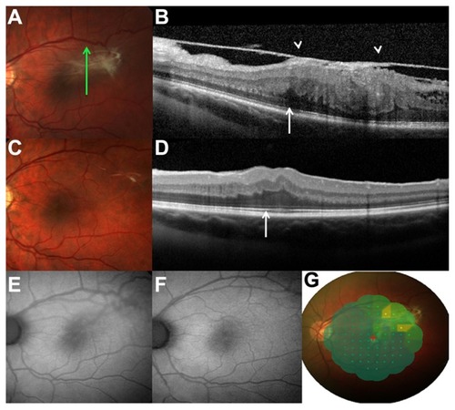
Figure 1 (A) Photograph of the fundus of the right eye in an 18-year-old girl who complained of metamorphopsia and reduced visual acuity, which was due to retinal folding corresponding to the epiretinal membrane underlying CHR-RPE in the macula. (B) Photograph during preoperative fundus examination of a 52-year-old man affected by CHR-RPE, showing a slight elevation on the optic disc and macula, retinal vessel tortuosity, hyperpigmentation, and extensive epiretinal membrane. (C) Photograph of preoperative peripheral CHR-RPE with epiretinal membrane in a 33-year-old man, showing retinal vessel tortuosity and visual distortion.
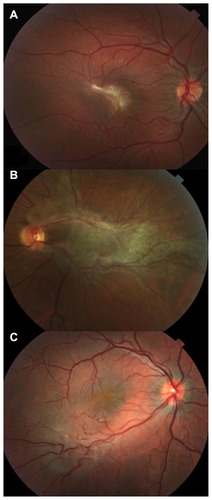
Figure 2 (A) Photograph during the preoperative fundus examination of a 26-year-old man, with slight elevation at the temporal macula, and hyperpigmentation and epiretinal membrane with retinal vessel tortuosity. (B) Preoperative spectral domain optical coherence tomography analysis showing disorganization of the normal retinal layers, loss of inner-outer segment photoreceptor junction (arrow), an elevated lesion with high reflectivity of the inner retina, hyporeflective shadowing of the tissue, and epiretinal membrane with vitreous traction (arrowheads). (C) Photograph 4 years postoperatively, where the epiretinal membrane has disappeared and the retinal vessel tortuosity has been relieved. (D) Spectral domain optical coherence tomography analysis at 4 years shows a normal appearance of the retinal layers and a clear inner-outer segment photoreceptor junction. (E) Preoperative fundus autofluorescence demonstrates a block in the background autofluorescence at the site of the lesion, and a slightly hyperautofluorescent area with the slightly indistinct appearance of the macular edema and epiretinal membrane. (F) At 4 years after surgery, fundus autofluorescence imaging detected normal autofluorescence of the macular region, with a reduction in retinal vessel tortuosity. (G) MP-1 highlighting lower retinal sensitivity corresponding to the area with higher adherence of the epiretinal membrane to the combined hamartoma of the retina and retinal pigment epithelium lesion preoperatively.