Figures & data
Figure 1 (a) Digital slit-lamp image of the right eye of the patient depicting diffuse conjunctival congestion, 8×8 mm creamy white full-thickness infiltrate (black arrowhead), stromal melt and nasal peripheral furrowing (white arrowhead) with guttering along with 3 mm anterior chamber hypopyon (severe ulcer). (b) Digital slit-lamp image of the right eye of the patient depicting diffuse conjunctival congestion, crescentic eccentric 7×7 mm mid to posterior stromal infiltrate (black arrowhead), inferonasal full-thickness infiltrate, nasal limbal spread (white arrowhead), and central corneal thinning with impending perforation (severe ulcer).
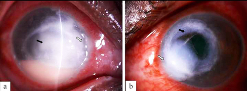
Figure 2 (a) Digital slit-lamp image of the patient depicting diffuse conjunctival congestion, superficial vascularization, total full-thickness corneal infiltrate with 360-degree limbal infiltrate and paralimbal thinning (black arrowhead) with central impending perforation (white arrowhead) (severe ulcer). (b) Digital slit-lamp image of the patient depicting diffuse conjunctival congestion, total graft infiltrate (black arrowhead), graft host junction melt from 1 to 7 o’clock (white arrowhead) and loose sutures along with superficial vascularization.
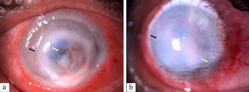
Table 1 Proposed Modified Jones Criteria for Pythium insidiosum Keratitis (Gurnani and Kaur Severity Grading of Pythium insidiosum Keratitis)Citation3,Citation5,Citation11
Table 2 Hallmark Clinical Features of Pythium insidiosum Keratitis and Its Masqueraders
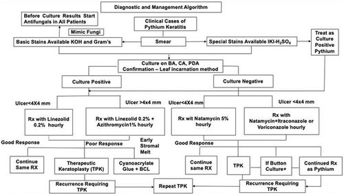
Figure 3 The novel diagnostic and management protocol of Pythium insidiosum keratitis.