
Figures & data
Figure 1 Flow chart describing study population. Deceased subjects and patients with missing data (n=10) were excluded from the one-year study population, resulting in a study population (N=103) with a four-year outcome.
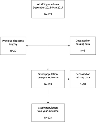
Table 1 Demographic Description for Study Population with Four-Year Outcome
Table 2 Outcome with Different Intraocular Pressure (IOP) Targets
Figure 2 Survival plot based on Kaplan–Meier estimates showing four-year outcome of XEN implantation (N=103) and probability of clinical success, the remaining cases with complete success (target IOP attained without pharmacologic treatment) and partial success (target IOP attained with the aid of pharmacologic agents), a total of 45 cases (43.7%) after 48 postoperative months.
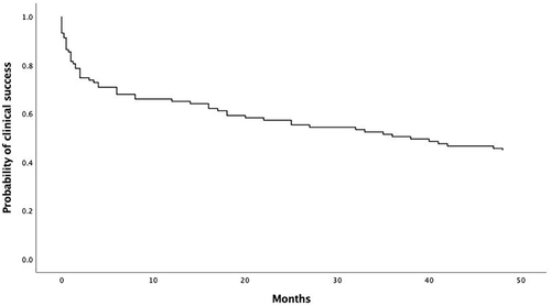
Figure 3 Pharmacologic treatment of glaucoma patients subjected to XEN implantation, showing mean number of topical substances and mean intraocular pressure (IOP) preoperatively and after four postoperative years. All XEN-patients in the cohort (N=103) were included initially and patients with secondary glaucoma surgery were excluded at the time of the secondary surgery.
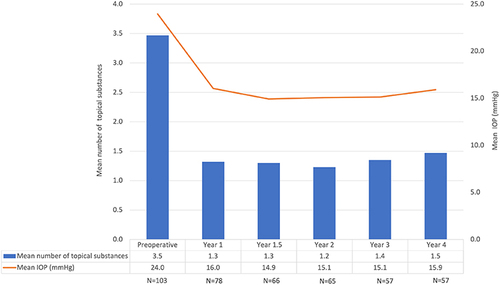
Figure 4 Survival plot based on Kaplan–Meier estimates for XEN implantation with different types of glaucoma; primary open-angle glaucoma (n=48) and pseudoexfoliative glaucoma (n=41). The probability of clinical success was 43.8% and 43.9% respectively after 48 months follow-up. Cox proportional hazard ratio=0.89 (95% CI=0.51–1.55), p=0.68.
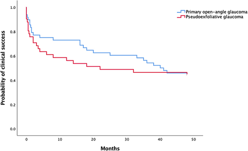
Figure 5 Survival plot based on Kaplan–Meier estimates for XEN gel stent procedure when combining XEN and cataract, with phacoemulsification and intraocular lens implantation (n=12), and for XEN stand-alone (n=91). Probability of clinical success was 50.0% and 42.9% respectively after 48 months follow-up. Cox proportional hazard ratio=1.38 (95% CI=0.59–3.20), p=0.46.
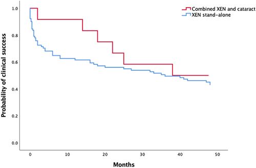
Table 3 Stent misplacement and Final Failure Rates
Figure 6 Survival plot based on Kaplan–Meier estimates for XEN gel stent procedures for different surgeons (A–D). Probability of clinical success was 37.5% for surgeon A (N=16), 55.7% for surgeon B (N=61), 16.7% for surgeon C (N=18) and 25.0% for surgeon D (N=8), after 48 months follow-up.
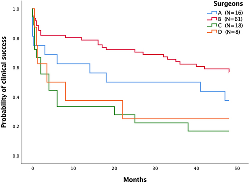
Table 4 Complication Rates
Figure 7 (A) January 2016: functioning but curved XEN 45 gel stent in place. IOP 18 mmHg without treatment 15 days after surgery. (B) March 2018 prior to needling. Pigment in XEN stent lumen, no effusion, IOP 21 mmHg on brinzolamide/brimonidine tartrate (Simbrinza®), bimatoprost (Lumigan®) and betaxolol (Betoptic®). The stent has migrated out of the anterior chamber and has no visible connection with aqueous humor on gonioscopy. (C) March 2018 post needling. The Xen stent lumen has been massaged through the intact conjunctiva towards the anterior chamber and the stent lumen is visible again on gonioscopy. Needling under and above the outer stent ostium has been performed straightening the stent, with no initial effect om filtration. The utmost part of the stent with pigmented embolus has thereafter been cut by needling (arrow), pigment and aqueous humor have drained to the subconjunctival space forming an ordinary filtration bleb (asterisk). IOP after one week washout of antihypertensive drugs = 12 mmHg.
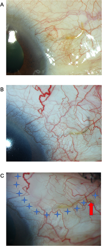
Table 5 Comparison of Our Data at Four Years and Some of the Largest Studies with at Least Three Years of Follow-Up