
Figures & data
Figure 1 (A) The 6mm × 6mm Angio en face OCT-A image centered on fovea. (B) The 15mm × 15mm Montage en face OCT-A whole retina image.
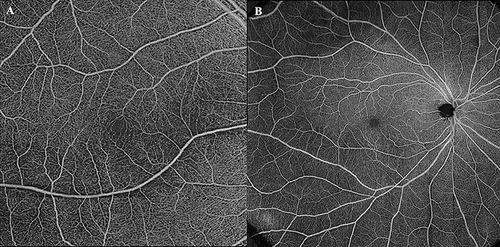
Figure 2 The 6mm × 6mm OCT-A image of a BRAO patient is divided into 9 ETDRS subfields. (A) Superficial vessel heat map. (B) Deep vessel heat map. (C) Macular thickness map. (D) Choroidal thickness map.
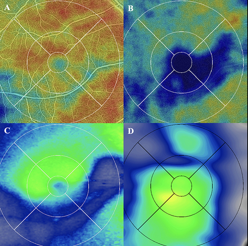
Table 1 Patient Demographic Information
Table 2 Correlation Between Retinal Thickness, SCP VD, DCP VD, Choroidal Thickness, Choroidal Volume vs Vision in RAO Eyes and Contralateral Eyes