Figures & data
Figure 1 Preoperative photograph, computer tomography, and postoperative photograph of the left eye. (A) Preoperative photography of left eye shows corneal laceration with corneal edema, the traumatic cataract, and tip of an intraocular foreign body (white arrow). (B) Computer tomography image shows the intraocular foreign body (white arrow) that penetrates into the vitreous via the lens. (C) Postoperative photography on day 3 after the operation shows improved corneal edema.
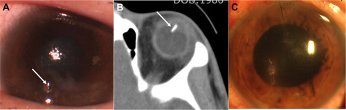
Figure 2 Slit-lamp photography of the superior retina by examination with a 90 diopter Volk lens. (A–C) Slit-lamp photography of the superior retina by examination with a 90 diopter Volk lens shows retinal necrosis spreading circumferentially in the periphery.
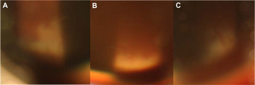
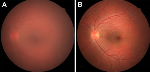
Figure 3 Fundus photography 3 days and 1 month after the operation. (A) Three days after the operation, fundus photography shows vitreous haziness due to inflammation. (B) Inflammation was resolved at 1 month after the operation.