Figures & data
Figure 1 Side profile of zig-square wound created by the femtosecond laser.
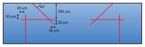
Table 1 Subject demographics, incisions dimensions and suturing technique
Table 2 Visual acuity preoperatively and at postoperative month 12
Table 3 Manifest and Sim K astigmatism preoperatively and at postoperative month 12
Table 4 Endothelial cell density preoperatively and at postoperative month 12
Figure 2 Photographs and Optovue® anterior segment optical coherence tomographic image from patient 5. (A) Preoperative photograph. (B) Postoperative photograph at week 8. (C) One year after femtosecond laser-assisted penetrating keratoplasty, there was a smooth anterior surface and good wound healing and apposition of the interlocking zig-square configuration.
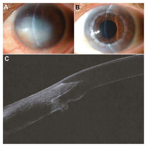
Figure 3 Optovue® anterior segment optical coherence tomographic image of patient 9 at 6 months after femtosecond laser-assisted penetrating keratoplasty, showing a well apposed donor-recipient junction with the zig-square incision.
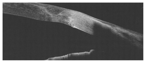
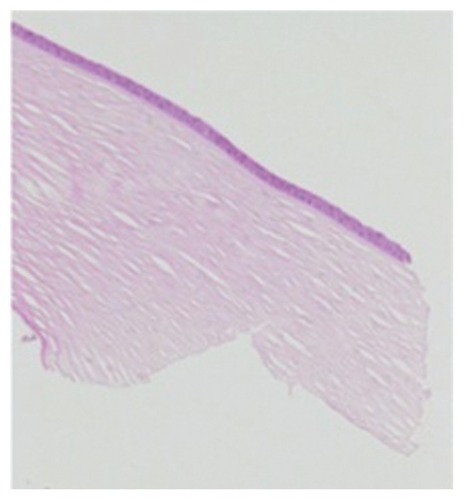
Figure 4 Light microscopy of the donor corneal rim (hematoxylin and eosin, 40×), demonstrating precise incision by the femtosecond laser, with minimal disruption to the surrounding architecture.