
Figures & data
Figure 1 Illustration of step-by-step surgical procedures. (a) Conjunctival peritomy. (b) An infusion cannula is inserted at infero-temporal and 4 scleral markings are made. (c) 6 mm scleral tunnel incision at superior sclera. (d) Internal corneal incision connecting the tunnel and anterior chamber. (e) Polypropylene 8–0 sutures are inserted into each lens eyelet. (f) IOL is positioned in a correct intraocular position at the entrance of the tunnel and each thread is pulled and externalized from each sclerotomy using a micro-forceps. (g) IOL is inserted and the suture is tightened evenly until the IOL is central. (h) The sutures are tied, and the knots are rotated and internalized through the sclerotomy sites. (i) Suture the scleral tunnel. (j) Suture the conjunctiva.
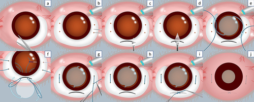
Figure 2 Complete surgical procedures. (a) Lidocaine subconjunctival injection. (b) Sub-tenon block. (c) 270-degree conjunctival peritomy. (d) An infusion cannula is inserted at infero-temporal. (e) 2 scleral markings at the temporal side adjacent to the 3 o’clock position. (f) 2 scleral markings at the nasal side adjacent to 9 o’clock position, 2 mm from the limbus and 4 mm apart between each marking (2 mm superior and 2 mm inferior to 3 and 9 o’clock points). (g) 6 mm scleral tunnel incision created at superior sclera using slit knife. (h) The lamellar sclerocorneal tunnel was made using crescent knife. (i) Internal corneal incision connecting the tunnel and anterior chamber. (j) IOL was evacuated. (k) Complete vitrectomy and vitreous shaving are performed. (l) Ophthalmic viscoelastic device (OVD) was injected. (m) Polypropylene 8-0 sutures inserted into each lens eyelet, IOL is positioned in a correct intraocular position at the entrance of the tunnel. (n) Nasal side thread is pulled and externalized from each sclerotomy using micro-forceps. (o) Temporal side thread is also pulled and externalized and IOL is then inserted. (p) Stabilize the IOL. (q) The suture is tightened evenly until the IOL is central. (r) The knots are rotated and internalized through the sclerotomy sites. (s) The scleral tunnel is sutured with 8-0 vicryl. (t) Conjunctiva is sutured using 8-0 426443 vicryl. (u) Antibiotic subconjunctival injection. (v) Surgery procedures are done.
