
Figures & data
Figure 1 The optic nerve and peri-papillary area; (A) OCTA and segmentation into superior inferior nasal and temporal quadrants, (B) disc photography, (C) peri-papillary OCT retinal nerve fiber layer thickness.
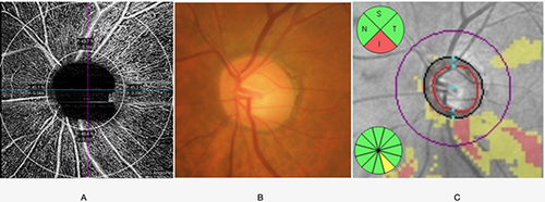
Table 1 Demographic and Clinical Characteristics of Subjects in the Healthy, Glaucoma Suspected and Glaucomatous Groups
Table 2 Ocular Parameters in Healthy, Glaucoma Suspected and Glaucomatous Groups and in Glaucomatous Eyes Differing by Severity (Adjusted for Age)
Figure 2 OCTA results in healthy (A), glaucoma suspected (B), and glaucomatous eyes (C).
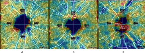
Figure 3 OCTA results in early (A), moderate (B), and severe glaucomatous eyes (C).
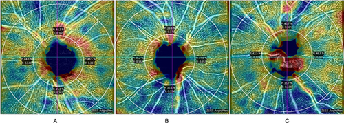
Table 3 Correlations Among Vessel Perfusion Density, Flux Index, Optical Coherence Tomography Retinal Nerve Fiber Layer Thickness (OCT RNFL), Optical Coherence Tomography Ganglion Cell Complex (OCT GCC) and Humphrey Visual Field Test 24–2 (HVF 24–2)
Table 4 Rates of Change per Year in Vessel Perfusion Density, Flux Index, Retinal Nerve Fiber Layer Thickness, Ganglion Cell Complex and Humphrey Visual Field 24–2 in Glaucomatous Eyes with and without Progression