
Figures & data
Figure 1 Comparison of objective refractions and subjective refraction measured under noncycloplegic condition. (A) Mean spherical equivalent (± standard deviation) measured by subjective method, SVS, RT7000, and Retinomax Screeen were −2.56 (3.00), −2.62 (2.38), −3.05 (2.84), and −3.26 (2.97), respectively. (B) J0 astigmatic component measured by subjective method, SVS, RT7000, and Retinomax Screeen were 0.08 (0.34), 0.03 (0.30), 0.12 (0.42), and 0.08 (0.42), respectively (C) J45 astigmatic component measured by subjective method, SVS, RT7000, and Retinomax Screeen were −0.02 (0.14), 0.03 (0.20), −0.04 (0.15), and −0.07 (0.18), respectively. *p < 0.05, **p < 0.01, ***p < 0.001.
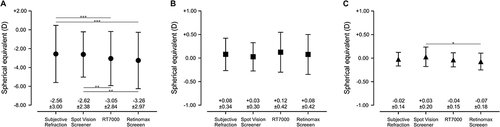
Figure 2 Comparison of objective refractions measured under cycloplegic condition and subjective refraction measured under noncycloplegic condition. Black symbols indicate results of non-cycloplegic condition and white symbols indicate results of cycloplegic condition. (A) Mean spherical equivalent (± standard deviation) measured by subjective method, SVS, RT7000, and Retinomax Screeen were −2.56 (3.00), −2.07 (2.66), −2.62 (2.98), and −2.66 (3.02), respectively. (B) J0 astigmatic component measured by subjective method, SVS, RT7000, and Retinomax Screeen were 0.08 (0.34), 0.05 (0.30), 0.13 (0.40), and 0.13 (0.41), respectively (C) J45 astigmatic component measured by subjective method, SVS, RT7000, and Retinomax Screeen were −0.02 (0.14), −0.02 (0.18), −0.04 (0.18), and −0.07 (0.19), respectively. ***p < 0.001.
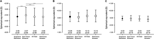
Figure 3 Difference in spherical equivalent between subjective refraction and three types of objective refraction under noncycloplegic condition visualized in Bland–Altman plots. (A) Bland–Altman plots of subjective refraction and objective refraction measured by Spot Vision Screener; Limits of agreement (LOA) was 0.05 ± 1.95. (B) Bland–Altman plots of subjective refraction and objective refraction measured by RT7000; LOA was 0.49 ± 0.97. (C) Bland–Altman plots of subjective refraction and objective refraction measured by Retinomax Screeen; LOA was 0.69 ± 1.39.
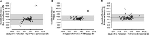
Figure 4 Difference in spherical equivalent between subjective refraction and three types of objective refraction under cycloplegic condition visualized in Bland–Altman plots. (A) Bland–Altman plots of subjective refraction and objective refraction measured by Spot Vision Screener; Limits of agreement (LOA) was −0.49 ± 1.25. (B) Bland–Altman plots of subjective refraction and objective refraction measured by RT7000; LOA was 0.06 ± 0.83. (C) Bland–Altman plots of subjective refraction and objective refraction measured by Retinomax Screeen; LOA was 0.10 ± 0.64.
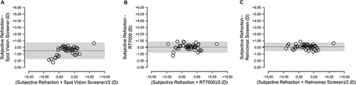
Table 1 Fixed and Proportional Errors for Objective Refractions When Compared to Subjective Refraction