Figures & data
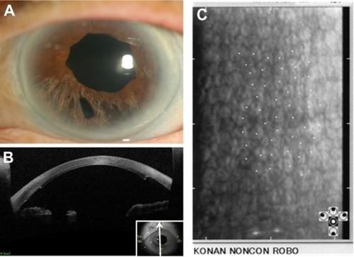
Figure 1 Slit lamp photographs, anterior segment optical coherence tomography (AS-OCT) image, and specular micrograph of the left eye before surgery. Slit lamp microscopy reveals iridoschisis in the inferior quadrant (A) and partial corneal edema in this portion (B). AS-OCT depicts a shallow anterior chamber with a separated anterior leaf of the iris touching the corneal endothelium (C). Specular microscopy of superior cornea shows endothelial cell loss (D).
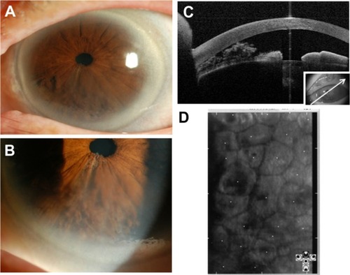
Figure 2 Slit lamp photographs of the left eye after cataract surgery with iridectomy. After the first surgery, slit lamp microscopy shows the implanted intraocular lens and partial removal of the iris in the lower quadrant (A). Corneal edema with Descemet’s folds are observed even in central corneal stroma (B).
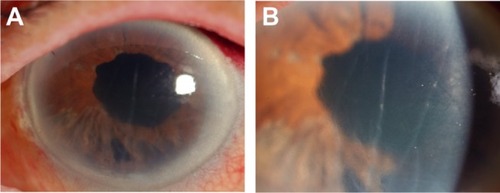
Figure 3 Slit lamp photograph, anterior segment optical coherence tomography (AS-OCT) image, and specular micrograph of the left eye after surgery. Slit lamp photograph at 6 months after nDSAEK shows that corneal edema has resolved (A). The pupil is slightly dilated with irregular margin due to sphincterotomy, and inferior iris atrophy is due to iridectomy. Other part of the iris is normal. AS-OCT image at 6 months post-nDSAEK shows adherence of the transplanted graft to recipient’s cornea, and iris defect due to iridectomy (B). Specular micrograph at 18 months post-nDSAEK shows high endothelial cell density in the center of the cornea (C).