Figures & data
Figure 1 The Alcon/WaveLight® FS200 patient interfaces 1504 (metal and glass, top) and 1505 (clear cone, bottom).
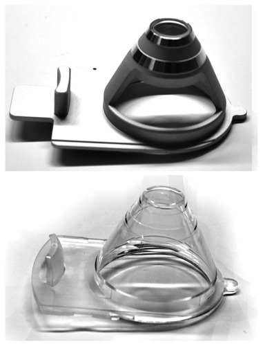
Table 1 Intended diameters of the flaps studied comparing the two groups
Figure 2 Measurement of flap thickness from the high-resolution meridional scan provided by the anterior-segment optical coherence tomography system.
Note: The specific flap was programmed to 120 μm of thickness.
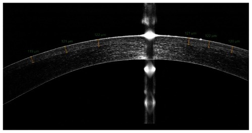
Figure 3 Flap diameter predictability using the clear cone interface 1505.
Notes: Vertical axis, measured difference in flap diameter = achieved postoperatively – programmed preoperatively. Horizontal axis, programmed flap diameter. All units in mm.
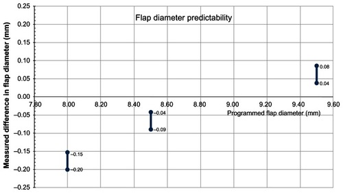
Figure 4 Flap thickness predictability using the clear cone interface 1505.
Notes: Measured central flap thickness (by anterior-segment optical coherence tomography imaging) versus programmed flap thickness. All units in μm.
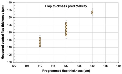
Table 2 Measured (via anterior-segment optical coherence tomography imaging) versus programmed flap thickness, as obtained using the clear cone interface