
Figures & data
Figure 1 Morphologic patterns of macular neovascularization (MNV) as visualized using swept-source OCT Angiography. Shape descriptors included (A) the “Medusa” pattern representing a lesion where vessels radiate in all directions; (B) the “Sea-fan” pattern representing a lesion where blood vessels radiate in all directions from one side; and (C) the “Tree-in-bud” pattern representing a round lesion with no obvious vascular trunk. Images were also evaluated for the presence of (D) peripheral loops (indicated by the yellow arrows), (E) capillary fringe (indicated by the yellow arrows), a perilesional dark halo (indicated by the box) and (F) a core vessel (indicated by the yellow arrows).
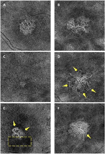
Table 1 Summary of Demographic and Clinical Characteristics for Eyes with MNV Secondary to Myopic Macular Degeneration (MMD) and Eyes with MNV Secondary to Age-Related Macular Degeneration (AMD)
Table 2 Summary of Qualitative and Quantitative Assessment of MNV Morphology Secondary to Myopic Macular Degeneration (MMD) and Age-Related Macular Degeneration (AMD) Using SS-OCTA
Table 3 Univariate Regression Analysis of MNV Area and Vessel Density
Table 4 Characteristics of Treatment-Naïve MNVs in Eyes with Discernible Lesions on En-Face SS-OCTA