Figures & data
Figure 1 (A) Photograph after Descemet stripping automated endothelial keratoplasty showing a white opacity (arrow) at the interface at the 12 o’clock position away from the temporal host corneal incision site. (B) Magnified view of the area of white opacity (arrow) at the interface. (C) Slit-lamp image showing epithelial ingrowth (arrow) between the graft and the host cornea. (D) Postoperative photograph after repeat Descemet stripping automated endothelial keratoplasty showing a clear cornea.
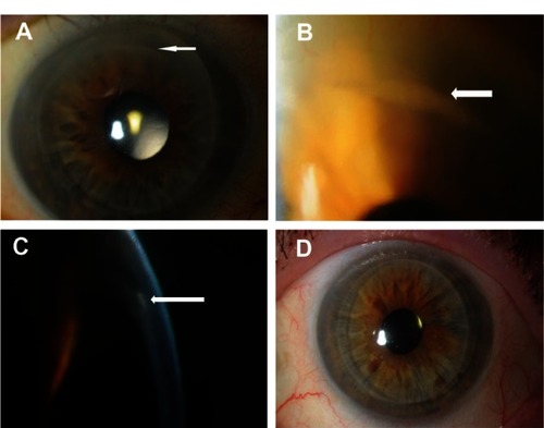
Figure 2 (A) Low power shot of the removed donor button to show full thickness of specimen at one side. Here there is epithelium (e) on part of the anterior face of the specimen. Also, Descemet’s membrane (d), is present on the back of the specimen, as well as, focally, at the front on this edge (hematoxylin and eosin, original magnification ×4). (B) Hematoxylin and eosin, original magnification ×40 to show epithelium along the anterior aspect of the specimen (arrow). Note that there is no Descemet’s membrane. (C) Hematoxylin and eosin, original magnification ×10 of the removed donor button shows view of area beneath epithelium. Arrow shows epithelium on the anterior face of the specimen. Posteriorly there is no endothelium. (D) High power of the back of the removed donor button specimen (hematoxylin and eosin, original magnification ×40). Shows graft Descemet membrane with some thickening and incipient guttae (arrow). There is no endothelium, suggestive of graft failure. Note the normal relationship of Descemet membrane to stroma.
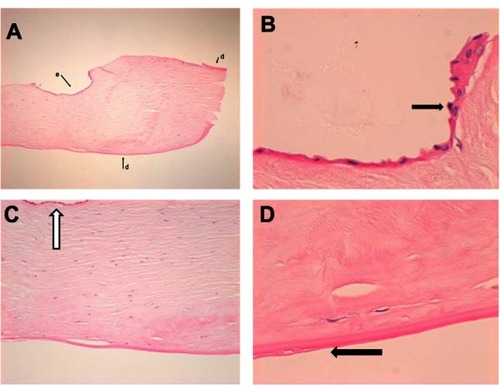
Table 1 Literature review of epithelial migration into anterior chamber after DSAEK