Figures & data
Figure 1 Patient with acute Stevens-Johnson syndrome, at the time of initial presentation to hospital and before initiation of treatment, showing conjunctival injection in both eyes.

Figure 2 Patient’s ophthalmic appearance at 2.5 days (60 hours) after disease onset. (A) The right eye demonstrates evidence of marked conjunctival injection. The right eye also shows defects in the bulbar conjunctival epithelium and epithelial sloughing of the eyelid margin. (B) The left eye shows similarly severe ophthalmic involvement.
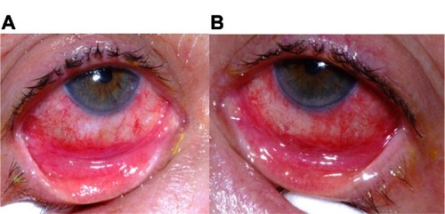
Figure 3 Right lower fornix of the patient five weeks after disease onset. Evident is a contracted tarsal conjunctival membrane (arrow) resulting in cicatricial entropion, trichiasis, and corneal epithelial breakdown.
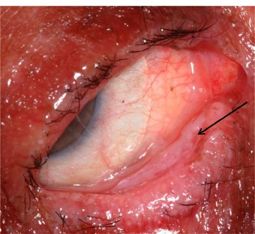
Figure 4 Patient showing a significant difference in clinical appearance of the right and left eyes four months after disease onset. (A) The right eye demonstrates a cicatricial entropion with associated trichiasis and conjunctival injection. A fluid-filled scleral lens (PROSE device) is in place. (B) The left eye demonstrates normal upper and lower eyelid positions without evidence of trichiasis. A PROSE device is also visible.
