Figures & data
Figure 1 Microscopy of host cornea showing the presence of a thin but distinct Descemet’s membrane (arrow). The specimen was stained with periodic acid-Schiff.
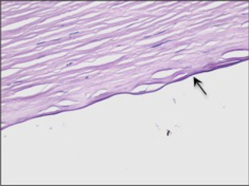
Figure 2 In vivo confocal microscopy enables high resolution imaging of the cornea and conjunctiva. This image reveals a clear image of the corneal endothelium with healthy cell count and thickness of 502 microns.
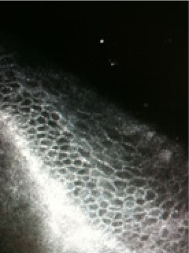
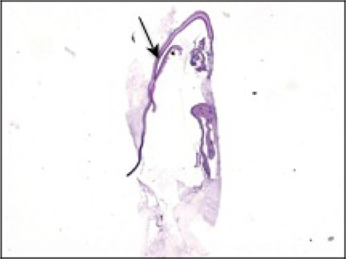
Figure 3 Microscopy of retained Descemet’s membrane (arrow) stained with periodic acid-Schiff.