
Figures & data
Figure 1 Visualization of residual ophthalmic viscosurgical device. (a) The UV light source and digital camera. The UV light source was illuminated from the side of the posterior pole. The porcine eye was photographed from the posterior pole side using a digital camera. (b) A representative image that shows fluorescein in the residual ophthalmic viscosurgical device visualized under UV light (white arrows).
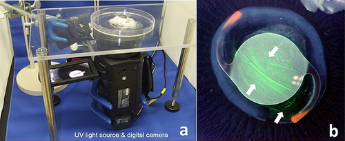
Figure 2 Intracapsular residual OVD under UV light (white arrows) visualized using fluorescent Si in various IOLs.
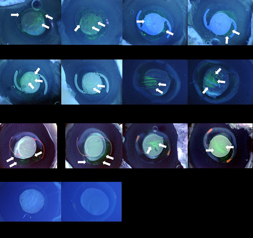
Table 1 Ophthalmic Viscosurgical Device
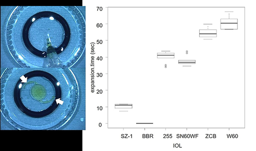
Figure 3 Two O-rings, each with a diameter of 13 mm and a height of 2 mm, were stacked to form a pseudo-lens capsule. The IOL was inserted, and the time required for the haptics to reach the inner wall of the ring (white arrows) was measured. The expansion time for each IOL is shown (right). The order of IOLs is from right to left in the decreasing order of the amount of residual OVD.
Data Sharing Statement
The datasets generated during the current study are not publicly available but are available from the corresponding author on reasonable request.