
Figures & data
Figure 1 The frequency of occurrence of individual ophthalmological symptoms in COVID-19 patients who developed ophthalmological symptoms, based on a 2021 study of 7300 individuals, representing 11.03% of these patients.Citation24
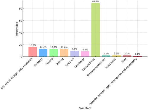
Figure 2 Changes observed in the retina based on the SERPICO-19 study published in 2020.Citation25
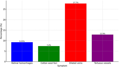
Figure 3 Central retinal vein occlusion in a 26-year-old female patient with Covid-19 infection and no other risk factors (Picture from the collection of the Ophthalmology Department, Medical University of Bialystok).
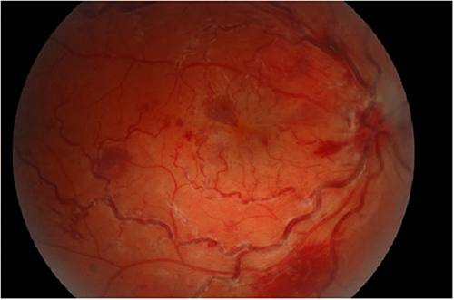
Figure 4 Paracentral acute middle maculopathy in a 56-year-old male patient with Covid-19 infection (Picture from the collection of the Ophthalmology Department, Medical University of Bialystok).
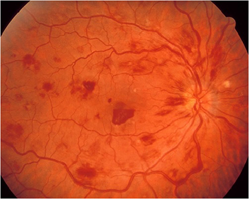
Figure 5 Uveitis during COVID-19 in a 48-year-old female patient (Picture from the collection of the Ophthalmology Department, Medical University of Bialystok).
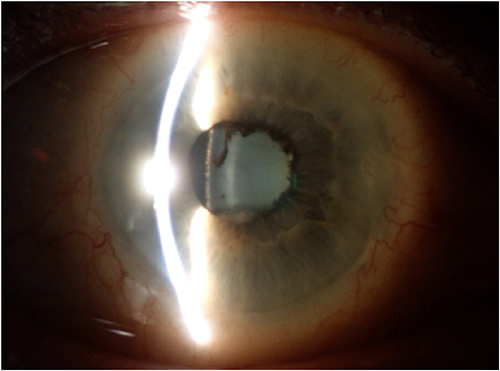
Figure 6 Bilateral optic neuritis associated with Covid-19 disease with no evidence of infection other than SARS-CoV-2 virus in a 45-year-old male patient. (A) right eye, (B) left eye (Picture from the collection of the Ophthalmology Department, Medical University of Bialystok).
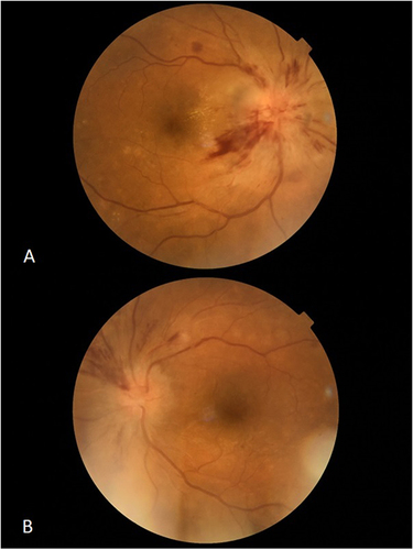
Table 1 Type of Ophthalmic Pathology in COVID-19 Disease Depending on the Location