Figures & data
Table 1 Demographics of Patients
Table 2 The Visual Acuity and Electroretinographic Parameters Before and After Silicone Oil Removal
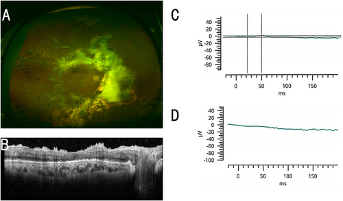
Figure 1 Findings in a 53-year-old woman who had ERG responses in her silicone (SO) filled eye. (A) Fundus photograph of the left eye after the SO removal. (B) Optical coherence tomographic (OCT) image of the left eye after the SO removal. (C) Combined rod-cone response before SO removal. (D) Combined rod-cone response after SO removal. The decimal visual acuity improved from 0.3 to 0.6 (from 0.52 to 0.22 logarithm of the minimum angle of resolution; logMAR units) after the SO removal.
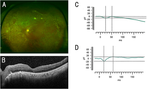
Figure 2 Representative ERGs of a 49-year-old man whose ERG was flat in the SO filled eye. (A) Fundus photograph of the right eye after the SO removal. (B) OCT image of the right eye after the SO removal. (C) Combined rod-cone responses before the SO removal. (D) Combined rod-cone responses after the SO removal. The decimal visual acuity improved from 0.2 to 0.4 (from 0.70 to 0.40 logarithm of the minimum angle of resolution; logMAR units) after the SO removal.