Figures & data
Figure 1 A 78-year-old man was treated with an intravitreal aflibercept injection. At baseline, the best-corrected visual acuity was 20/25. (A) Red-free photograph showing a pigment epithelial detachment, lipid, and serous retinal detachment at the macular area. (B) Fluorescein angiography image showing leakage due to occult choroidal neovascularization and a fibrovascular pigment epithelial detachment, as well as an occlusion of branch retinal vein and collaterals at the upper macula. (C and D) Early-phase (left) and late-phase (right) indocyanine green angiography showing no polypoidal lesion. (E and F) Baseline horizontal (left) and vertical (right) optical coherence tomography (Spectralis®; Heidelberg Engineering, Heidelberg, Germany) images showing a serous retinal detachment and fibrovascular pigment epithelial detachment.
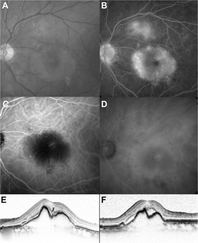
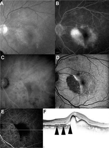
Figure 2 A month after the first intravitreal aflibercept injection. The best-corrected visual acuity was 20/28. (A) Red-free photograph showing an area of retinal pigment epithelium defect inferior to the fovea. (B) Fluorescein angiography image showing an area of hypofluorescence. (C) Late-phase indocyanine green angiography showing hypofluorescence. (D) Fundus autofluorescence showing an area of hypo autofluorescence. (E) Early-phase indocyanine green angiography and horizontal optical coherence tomography image. (F) Scanned by the white arrow on the left indocyanine green angiography image, which was simultaneously obtained by spectral-domain optical coherence tomography, showing defect of the retinal pigment epithelium line (arrows).