Figures & data
Figure 1 Initial anterior segment findings of the right eye. Central corneal opacity, corneal thinning, and iridocorneal adhesion were observed.
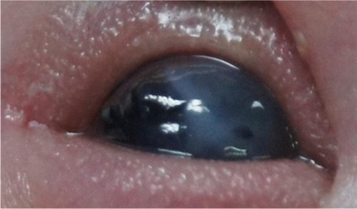
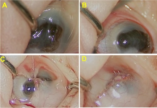
Figure 2 Intraoperative findings. Iris protrusion and a fat anterior chamber were observed.
Notes: (A) A 360° peritomy was performed to increase the conjunctival mobility. (B) The flap was brought down to cover the cornea. Simple interrupted sutures with 8–0 Vicryl were used (C and D).