Figures & data
Figure 1 Fundus photographs of the right eye (A and B) and left eye (C and D), and photographs focused on the optic disc of the right eye (E and F) and left eye (G and H) taken in 2009 and 2012, respectively. spectral-domain optical coherence tomography scans of the optic disc in the right eye (I) and left eye (J) in 2012. (A–D) Fundus photographs show obvious bilateral optic disc swelling, and absence of vitreous opacifications and retinal exudates. (E and F) Fundus photographs focused on the optic disc reveal the lack of a sharp outline to the redness of the optic discs, and retinal vascular sheathing around the optic discs. There are no dilated superficial capillaries and hemorrhages on optic discs as was observed in 2009. (G and H) Both optic discs were white in color and retinal vascular sheathing was increased after 3 years. (I and J) Longitudinal scans of the optic discs showing obvious optic disc swelling.
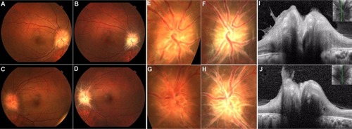
Figure 2 Slit-lamp examinations of the right eye (A) and left eye (B) in 2012. slit-lamp biomicroscopy revealed strong conjunctivitis and episcleritis in both eyes treated with mydriatics.
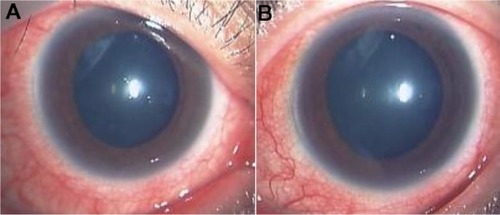
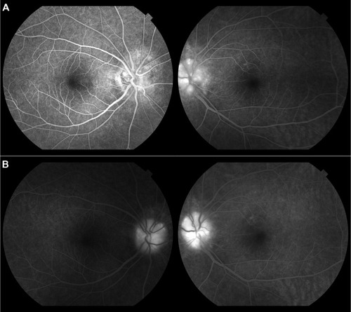
Figure 3 Fluorescein angiography at early phase (A) and late phase (B) in 2012. Fluorescein angiography showed strong staining and weak late phase leakage from the optic disc in both eyes. There were no dilated superficial capillaries on optic discs.