Figures & data
Figure 1 (A) Vertical EDI OCT and (B) HP-OCT images of the deep optic nerve head and deep peripapillary structures.
Notes: The anterior (down arrow) and posterior (up arrow) borders of the lamina cribrosa are identified as points at which the highly reflective lamina cribrosa starts and ends, respectively. The EDI OCT image shows a sharper contrast between the lamina cribrosa and the surrounding structures. The signal strength of the sclera (*) is stronger in the HP-OCT image than in the EDI OCT image. Scale bar = 200 μm.
Abbreviations: EDI OCT, enhanced-depth imaging optical coherence tomography; HP-OCT, high-penetration optical coherence tomography.
Abbreviations: EDI OCT, enhanced-depth imaging optical coherence tomography; HP-OCT, high-penetration optical coherence tomography.
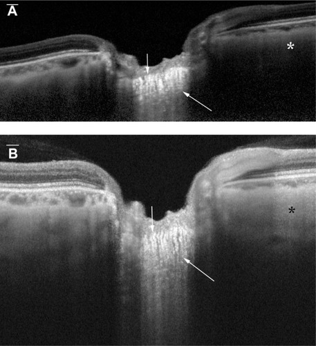
Figure 2 Representative HP-OCT image showing scleral vessels.
Notes: The vertical HP-OCT image shows the intrascleral path of vessels (arrows). The vessels are seen as continuous hyporeflective areas surrounded by the hyperreflective sclera.
Abbreviation: HP-OCT, high-penetration optical coherence tomography.
Abbreviation: HP-OCT, high-penetration optical coherence tomography.
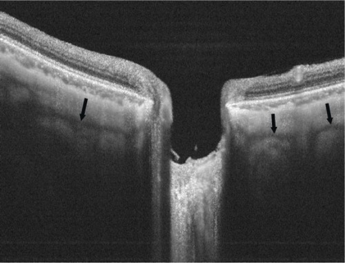
Table 1 Mean subjective visibility scores with EDI OCT and HP-OCT, for deep the ONH and parapapillary structures
Figure 3 Mean visibility scores of the deep peripapillary structures, by quadrant.
Notes: The subjective visibility scores of the intrascleral vessels (♦) with EDI OCT (P < 0.0001), HP-OCT (P = 0.0009), and overall (P < 0.0001). Visibility of the cerebrospinal fluid space (■) with EDI OCT (P < 0.0001), HP-OCT (P < 0.0001), and overall (P < 0.0001). Visibility of the choroid (▲) with EDI OCT (P < 0.0001), HP-OCT (P = 0.3350), and overall (P < 0.0001). The best mean visibility scores were in the temporal quadrant images, with both EDI OCT and HP-OCT.
Abbreviations: CSF, cerebrospinal fluid; EDI OCT, enhanced-depth imaging optical coherence tomography; HP-OCT, high-penetration optical coherence tomography.
Abbreviations: CSF, cerebrospinal fluid; EDI OCT, enhanced-depth imaging optical coherence tomography; HP-OCT, high-penetration optical coherence tomography.
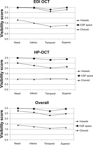
Table 2 Mean visibility scores of the deep ONH structures in horizontal and vertical images using EDI OCT, HP-OCT, and overall