Figures & data
Table 1 Demographic data and clinical outcomes of two patients (three eyes) with microcornea
Figure 1 Surgical technique of non-Descemet’s stripping automated endothelial keratoplasty in case 2. (A) Under the surgical microscope, the recipient corneal size was horizontal 9.0 mm × vertical 7.5 mm, and the donor diameter was determined as 6.0 mm. (B) A 6.0 mm diameter donor lamella, in which the endothelial surface was coated with viscoelastic material, was loaded into the Busin glide. (C) Without recipient descemetorhexis, the donor lamella was easily inserted into the anterior chamber with a pull-through technique using the sheets glide and the Busin glide. Note the violet dot marking was 8.0 mm in diameter. (D) after securing the wound with an interrupted 10-0 nylon suture, air was injected to press the donor tissue against the recipient cornea.
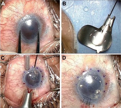
Figure 2 Slit-lamp photographs and anterior segment optical coherent tomography images in case 3. (A and B) Preoperatively, mild stromal edema and shallow anterior chamber were noted, although best-corrected visual acuity was 20/20. A flat corneal section was noted by anterior segment optical coherent tomography. (C and D) At one day postoperatively, partial donor detachment (arrow) was noted, and rebubbling was performed immediately. (E and F) At 2 months postoperatively, total donor attachment was noted.
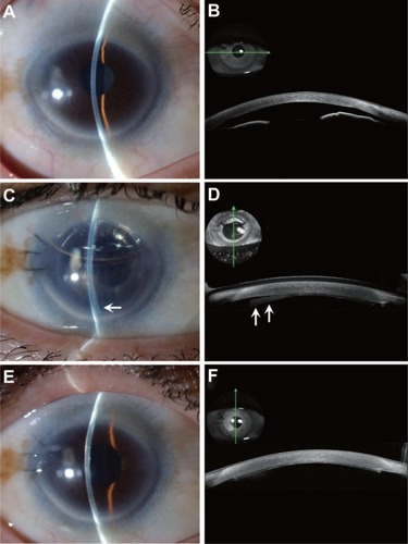
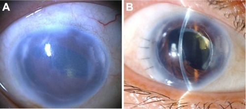
Figure 3 Slit-lamp photographs in case 1 with microcornea and iridocorneal endothelial syndrome. (A) Preoperatively, severe corneal edema, pupil deformity, and near 360° peripheral anterior synechia were noted. (B) Postoperatively, the cornea became clear. Best-corrected visual acuity improved from 20/1000 to 20/100.