42
Views
0
CrossRef citations to date
0
Altmetric
Case Series

Delayed macular hole formation after demarcation laser photocoagulation for subclinical retinal detachment

, , &
Pages 915-917 | Published online: 13 May 2014

Figures & data

Figure 1 (A) Wide field fundus photograph showing a large horseshoe tear in the superotemporal area after demarcation laser photocoagulation barricade. (B) Time domain optical coherence tomography of the left eye showing a full thickness macular hole with an epiretinal membrane. (C) Time domain optical coherence tomography of the left eye showing regression of the macular anatomy to normal after macular hole repair.

Figure 1 (A) Wide field fundus photograph showing a large horseshoe tear in the superotemporal area after demarcation laser photocoagulation barricade. (B) Time domain optical coherence tomography of the left eye showing a full thickness macular hole with an epiretinal membrane. (C) Time domain optical coherence tomography of the left eye showing regression of the macular anatomy to normal after macular hole repair.

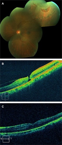
Table 1 Patient characteristics