Figures & data
Figure 1 Corneal clearance on slit lamp biomicroscopy.
Note: Scleral lens (white arrow) with haptic resting on sclera and the space between the scleral lens and the cornea – corneal clearance (black arrow).
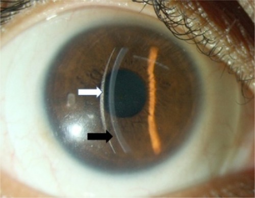
Table 1 Description of mini-scleral lenses available in the market
Figure 2 ASOCT in a patient with keratoconus with vault measured as 0.47 mm.
Note: The vault is more in periphery as compared to the center.
Abbreviation: ASOCT, anterior segment optical coherence tomography.
Abbreviation: ASOCT, anterior segment optical coherence tomography.
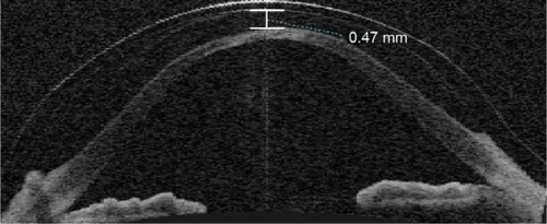
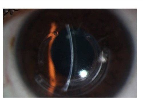
Figure 3 PROSE in keratoconus.
Notes: Scleral lens in keratoconus patient having intracorneal rings. The patient was referred for keratoplasty as he was not able to wear RGP lenses. With PROSE having FSE of 0.6, his visual acuity improved to 20/20.
Abbreviations: FSE, front surface eccentricity; PROSE, prosthetic replacement of the ocular surface ecosystem; RGP, rigid gas permeable.
Abbreviations: FSE, front surface eccentricity; PROSE, prosthetic replacement of the ocular surface ecosystem; RGP, rigid gas permeable.