Figures & data
Figure 1 (A and B) Fundus photographs showing yellowish, oval-shaped drusenoid-like lesion with attenuation of the foveal reflex in both eyes. (C and D) Red-free fundus photographs showing heterogeneous foveal lesions. (E and F) Fluorescein angiogram showing early foveal hyperfluorescence with late ill-defined leakage in right eye and left eye, respectively. (G and H) Autofluorescence photos showed heterogeneous hyperfluorescence in the macula of both eyes.
Note: A,C,E, and G are right eye; B,D,F, and H are left eye.
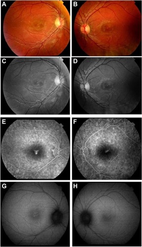
Figure 2 Optical coherence tomography. (A and B) Disruption of the outer retinal layers with nonspecific retinal thickening in both eyes at presentation. (C and D) Disruption of the inner segment/outer segment junction in both eyes, at 3 months after presentation.
Note: A and C are right eye; B and D are left eye.
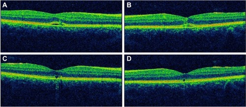
Table 1 Reported cases in the literature of retinal damage caused specifically by green lasers