
Figures & data
Figure 1 Patient 1.
Notes: Top: note the bilateral opacified, ectatic corneas, with protrusion of the globe and limbal staphylomas. Bottom left: UBM showing iris adherent to the posterior corneal surface (arrow) and absence of the crystalline lens (horizontal axial scan). Bottom right: T2 MRI bilateral large globes with dislocated lenses in the vitreous cavity (arrowheads). Dilation of the optic nerve sheath in the peripapillary region, suggestive of optic nerve hydrops, is noted.
Abbreviations: MRI, magnetic resonance imaging; UBM, ultrasound biomicroscopy.
Abbreviations: MRI, magnetic resonance imaging; UBM, ultrasound biomicroscopy.
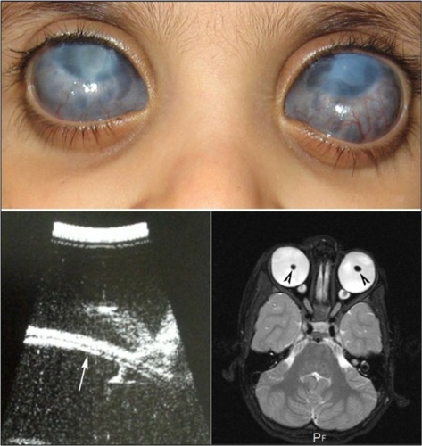
Figure 2 Patient 2.
Notes: Top: color photograph of the right eye, showing an ectatic vascularized cornea, with corneal/limbal staphylomas. Bottom left: UBM showing the iris adherent to the posterior corneal surface (arrow) and absence of the crystalline lens (radial scan with angle at 9 o’clock). Bottom right: T2 MRI showing bilateral enlarged globes with aphakia. Dilation of the optic nerve sheath in the peripapillary region, suggestive of optic nerve hydrops, is noted.
Abbreviations: MRI, magnetic resonance imaging; UBM, ultrasound biomicroscopy.
Abbreviations: MRI, magnetic resonance imaging; UBM, ultrasound biomicroscopy.
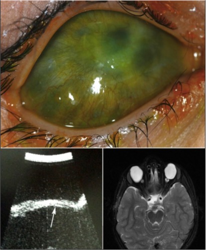
Table 1 Clinical features and course of patients with congenital anterior staphylomas