
Figures & data
Table 1 Clinical and demographic data of the CRVO patients
Figure 1 Representative fundus color photograph, measurement of retinal thickness by OCT, and an ERG for the same eye.
Notes: (A) A fundus color photograph demonstrating CRVO in a 69-year-old woman. (B) A retinal thickness map obtained by OCT. (C) ERG waveforms (30 Hz flicker waveform on the left and cone waveform on the right) obtained with the le2000 ERG system. Tick marks on the X axis represent 10 ms intervals.
Abbreviations: CRVO, central retinal vein occlusion; ERG, electroretinogram; OCT, optical coherence tomography.
Abbreviations: CRVO, central retinal vein occlusion; ERG, electroretinogram; OCT, optical coherence tomography.
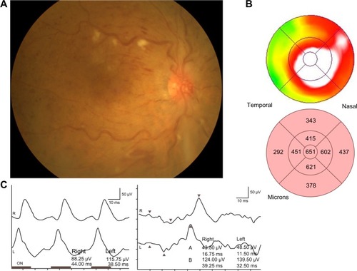
Table 2 Comparison of ERG parameters between the affected eyes and unaffected contralateral eyes of the CRVO patients
Table 3 Anatomic measurements in the CRVO patients with macular edema
Table 4 Association between ERG parameters and retinal thickness in the nine macular subfields
Table 5 Association between ERG parameters and retinal volume in the nine macular subfields
Table 6 Correlations between ERG parameters and the nonperfused area of the retina