Figures & data
Figure 1 (A) PAS diastase stain (×200) of the lesion confirming pink mucin within numerous ductal structures (arrow). (B) H&E stain (×200) of the lesion showing a predominantly well differentiated population of tubular ductal structures (arrow) and occasional squamoid cords extending beneath the conjunctival surface in mildly fibrotic stroma.
Abbreviations: H&E, hematoxylin and eosin; PAS, periodic acid-Schiff.
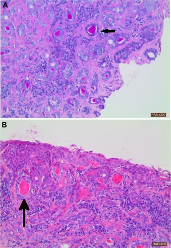
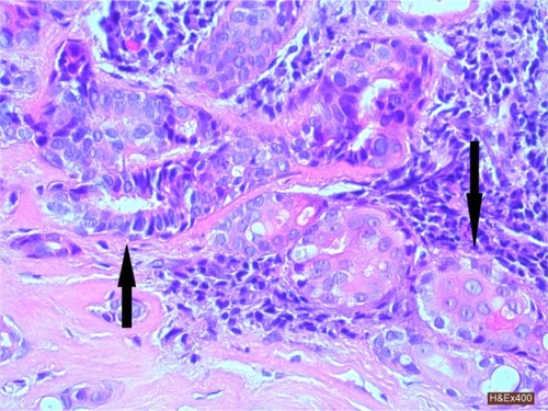
Figure 2 H&E stain (×400) of the tumor. Ductal structures lined by well differentiated columnar epithelium (up arrow) and solid squamoid nests with abundant eosinophilic cytoplasm (down arrow). There was minimal cytological atypia in either ductal or squamous component.
Abbreviation: H&E, hematoxylin and eosin.