69
Views
1
CrossRef citations to date
0
Altmetric
Case Series

Choroidal neovascularization secondary to Best’s vitelliform macular dystrophy in two siblings of a Malay family

, , , &
Pages 537-542 | Published online: 12 Mar 2014

Figures & data

Figure 1 (AH) Case 1. Color fundus photograph of the right eye at presentation, showing submacular hemorrhage and a yellowish fibrotic nodule (A). Color fundus photograph of the left eye, showing a dome-shaped elevation at the foveal region with the presence of yellowish deposits (B). Fundoscopy of the right eye, showing resolved subretinal hemorrhage with residual scarring at the foveal region at 2 months posttreatment (C). Fundoscopy of the left eye, showing similar findings to those at presentation (D). Fundus fluorescein angiogram, showing increased hyperfluorescence at the macula, consistent with choroidal neovascularization (E early phase, F late phase). Optical coherence tomography of the right eye at presentation, showing a subretinal pigment epithelium fibrotic nodule with a subretinal reflection of a subfoveal choroidal neovascularization and surrounding subretinal fluid (G). Optical coherence tomography of the right eye at 2 months posttreatment, showing resolution of the subretinal fluid at the right macula (H). Green lines indicate the cross-section of the macula shown by the adjacent optical coherence tomography image.

Figure 1 (A–H) Case 1. Color fundus photograph of the right eye at presentation, showing submacular hemorrhage and a yellowish fibrotic nodule (A). Color fundus photograph of the left eye, showing a dome-shaped elevation at the foveal region with the presence of yellowish deposits (B). Fundoscopy of the right eye, showing resolved subretinal hemorrhage with residual scarring at the foveal region at 2 months posttreatment (C). Fundoscopy of the left eye, showing similar findings to those at presentation (D). Fundus fluorescein angiogram, showing increased hyperfluorescence at the macula, consistent with choroidal neovascularization (E early phase, F late phase). Optical coherence tomography of the right eye at presentation, showing a subretinal pigment epithelium fibrotic nodule with a subretinal reflection of a subfoveal choroidal neovascularization and surrounding subretinal fluid (G). Optical coherence tomography of the right eye at 2 months posttreatment, showing resolution of the subretinal fluid at the right macula (H). Green lines indicate the cross-section of the macula shown by the adjacent optical coherence tomography image.

Figure 2 (AD) Case 2. Color fundus photograph of the right (A) and left (B) eyes showing dome-shaped elevation of the foveal regions, with the presence of yellowish deposits, signifying Best’s vitelliform macular dystrophy. Optical coherence tomography of the right (C) and left (D) eye revealed the presence of subretinal fluid at the foveal regions. Green lines indicate the cross-section of the macula shown by the adjacent optical coherence tomography image.

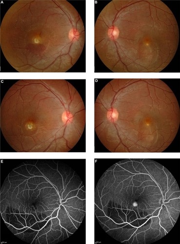
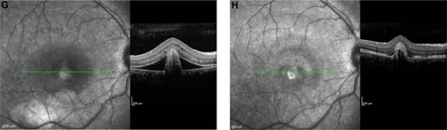
Figure 2 (A–D) Case 2. Color fundus photograph of the right (A) and left (B) eyes showing dome-shaped elevation of the foveal regions, with the presence of yellowish deposits, signifying Best’s vitelliform macular dystrophy. Optical coherence tomography of the right (C) and left (D) eye revealed the presence of subretinal fluid at the foveal regions. Green lines indicate the cross-section of the macula shown by the adjacent optical coherence tomography image.

Figure 3 (AD) Case 3. Color fundus photographs of the fourth child at the time of diagnosis of choroidal neovascularization (A and B). Right fundoscopy at 3 years posttreatment, showing a resolved subfoveal hemorrhage with a residual dome-shaped elevation and yellowish deposits at the foveal region (C). Left fundoscopy at 3 years postpresentation, showing an increase in the size of the dome-shaped elevation, with no sign of a submacular hemorrhage (D).

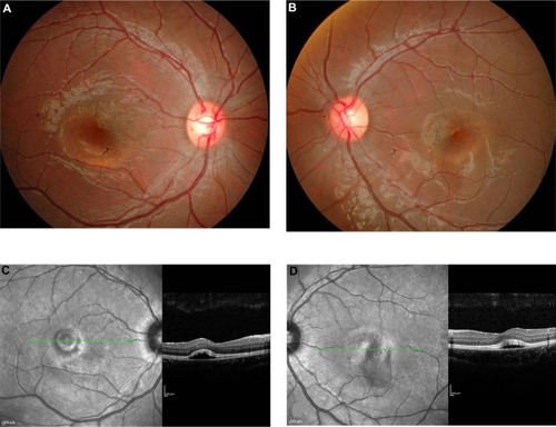
Figure 3 (A–D) Case 3. Color fundus photographs of the fourth child at the time of diagnosis of choroidal neovascularization (A and B). Right fundoscopy at 3 years posttreatment, showing a resolved subfoveal hemorrhage with a residual dome-shaped elevation and yellowish deposits at the foveal region (C). Left fundoscopy at 3 years postpresentation, showing an increase in the size of the dome-shaped elevation, with no sign of a submacular hemorrhage (D).

Table 1 Clinical summary