Figures & data
Figure 1 Fundus diagrams of retinal detachments at presentation and ocular parameters.
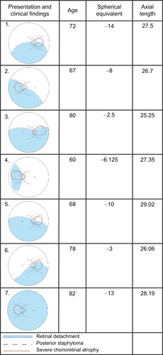
Table 1 Summary of clinical characteristics, management, and outcomes
Figure 2 Intraoperative photograph during drainage of subretinal fluid through juxtapapillary microhole in Case 4. Note proximity of drainage site to retinal vein.
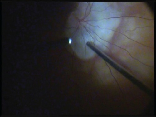
Figure 3 Preoperative optical coherence tomography image of Case 6 showing full-thickness juxtapapillary microhole (white arrow).
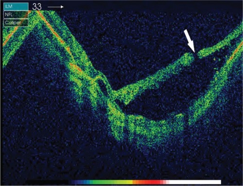
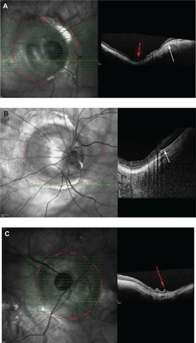
Figure 4 Postoperative optical coherence tomography images of (A) Case 7, (B) Case 4, and (C) Case 5 showing type 3 posterior staphyloma (outlined with dashed red line). Note retinal vascular folds (red arrows) and attenuated choroid and retinal pigment epithelium (white arrow).