Figures & data
Table 1 Clinical and imaging characteristics in three eyes with primary intraocular lymphoma
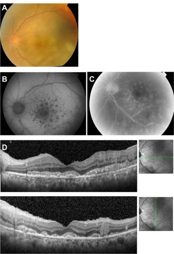
Figure 1 Case 1. Fundus photographs, fluorescein angiograms, and spectral-domain optical coherence tomographic images of the right eye in a 57-year-old man with primary intraocular lymphoma after vitrectomy.
Notes: (A) Color fundus photograph shows diffuse retinal infiltration in the macula and retinal vasculitis in the temporal and nasal areas. (B) Late phase fluorescein angiogram showing hypofluorescent spots with a leopard spot pattern in the posterior fundus and staining of the arteries and an avascular area in the nasal region. (C) Spectral-domain optical coherence tomographic images. (Left) Nodular hyper-reflective infiltration at the level of the retinal pigment epithelium, separation of Bruch’s membrane from the retinal pigment epithelium, partial damage of the retinal pigment epithelium, disruption of the photoreceptor inner segment/outer segment junction, and hyper-reflective signals in the inner retina can be seen. (Right) A layered structure of the retina cannot be detected on the temporal side.
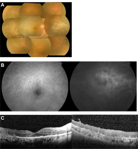
Figure 2 Case 2. Fundus images of the left eye in a 57-year-old man with primary intraocular lymphoma after vitrectomy.
Notes: (A) Color fundus photograph shows many small, yellowish lesions with distinct boundaries resembling drusen in the posterior fundus. (B) Cytological examination of vitrectomy specimen shows atypical lymphocytes with dyskaryosis and aberrant chromatin, stained using May-Grünwald-Giemsa. (C) Granular pattern of hypoautofluorescence and hyperautofluorescence is seen on fundus autofluorescence. (D) Late phase of fluorescein angiography shows hyperfluorescence of the disc and small spots with a reverse fluorescence pattern to the fundus autofluorescence pattern. (E) Spectral-domain optical coherence tomographic images. Nodular hyper-reflective infiltration at the level of retinal pigment epithelium and above the retinal pigment epithelium, a separation of Bruch’s membrane from the retinal pigment epithelium, partial destruction of the retinal pigment epithelium, disruption of the photoreceptor inner segment/outer segment junction line, and multiple hyper-reflective signals in the inner retina can be seen.
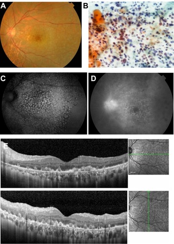
Figure 3 Case 3. Fundus images of the left eye in a 67-year-old man with primary intraocular lymphoma after vitrectomy.
Notes: (A) Fundus photograph showing many large whitish yellow spots with indistinct boundaries in the macular area. (B) Fundus autofluorescence image showing hypoautofluorescence and hyperautofluorescence surrounding a mass lesion with a honeycomb shape. (C) Late phase of fluorescein angiogram shows hyperfluorescence of the disc and fluorescence pattern which is the reverse of the fundus autofluorescence pattern. (D) Spectral-domain optical coherence tomographic image showing hyper-reflective infiltration at the level of the retinal pigment epithelium, separation of Bruch’s membrane from the retinal pigment epithelium, disruption of the photoreceptor inner segment/outer segment junction, hyper-reflective lesions in the form of bands above the retinal pigment epithelium, and hyper-reflective signals in the inner retina.